 Programmed death ligand 1 (PD-L1) is the principal ligand of the programmed death one (PD-1) receptor and is essential in the development of immune tolerance and prevention of autoimmunity. Unfortunately, PD-L1 is also expressed in many tumour types which is instrumental in allowing cancer cells to evade immune recognition. Immunohistochemistry may be used to quantify levels of PDL-1 and in turn may stratify patients and guide immunotherapy-based treatments. Unfortunately, current immunohistochemistry methods fail to capture much of the heterogeneity in PDL-1 expression that is necessary for guiding therapy. This has led to the need to develop advanced non-invasive imaging techniques which might quantify PD-L1 expression within individual lesions to guide future treatments and improve efficacy.
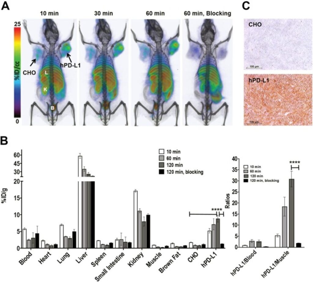
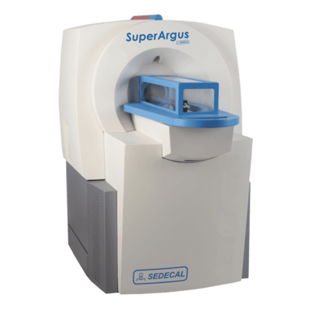
In this paper published in Molecular Imaging researchers from John Hopkins University developed a peptide-based, high-affinity PD-L1 imaging for positron emission tomography (PET) imaging. The radiotracer, [18F]FPy-WL12, was first assessed for specificity by flow cytometry and uptake of the radiotracer reflected the PD-L1 expression showing specificity of the tracer. In vivo evaluation of [18F]FPy-WL12 in mice bearing cancer xenografts was performed using the Sedecal SuperArgus PET/CT. In vivo PET/CT images demonstrated a significant PD-L1-specific uptake of [18F]FPy-WL12 in tumors. This significant uptake was reversed in mice receiving a blocking does of unmodified WL12, further confirming the specificity of the tracer
This study shows [18F]FPy-WL12 to be a specific radiotracer capable of quantifying heterogeneous distribution of PD-L1 in tumour models. Translation of this radiotracer into a clinical form would allow an accessible radiotracer specific to PD-L1 that is easily accessible and integrated into existing clinical workflows.
For more information about the Sedecal SuperArgus PET/CT system please reach out to Scintica Instrumentation.
Programmed death ligand 1 (PD-L1) is the principal ligand of the programmed death one (PD-1) receptor and is essential in the development of immune tolerance and prevention of autoimmunity. Unfortunately, PD-L1 is also expressed in many tumour types which is instrumental in allowing cancer cells to evade immune recognition. Immunohistochemistry may be used to quantify levels of PDL-1 and in turn may stratify patients and guide immunotherapy-based treatments. Unfortunately, current immunohistochemistry methods fail to capture much of the heterogeneity in PDL-1 expression that is necessary for guiding therapy. This has led to the need to develop advanced non-invasive imaging techniques which might quantify PD-L1 expression within individual lesions to guide future treatments and improve efficacy.
In this paper published in Molecular Imaging researchers from John Hopkins University developed a peptide-based, high-affinity PD-L1 imaging for positron emission tomography (PET) imaging. The radiotracer, [18F]FPy-WL12, was first assessed for specificity by flow cytometry and uptake of the radiotracer reflected the PD-L1 expression showing specificity of the tracer. In vivo evaluation of [18F]FPy-WL12 in mice bearing cancer xenografts was performed using the Sedecal SuperArgus PET/CT. In vivo PET/CT images demonstrated a significant PD-L1-specific uptake of [18F]FPy-WL12 in tumors. This significant uptake was reversed in mice receiving a blocking does of unmodified WL12, further confirming the specificity of the tracer
This study shows [18F]FPy-WL12 to be a specific radiotracer capable of quantifying heterogeneous distribution of PD-L1 in tumour models. Translation of this radiotracer into a clinical form would allow an accessible radiotracer specific to PD-L1 that is easily accessible and integrated into existing clinical workflows.
For more information about the Sedecal SuperArgus PET/CT system please reach out to Scintica Instrumentation.