RFLSI III Laser Speckle Perfusion Imager
Robust Blood Perfusion Imaging System

System Overview
RFLSI III is a blood perfusion imaging system that helps researchers to monitor and record, the blood perfusion of any exposed tissues or organs for microcirculation studies, in real-time, and to visualize the quantified data.
The RFLSS III Laser Speckle Imaging System is based on LSCI (Laser Speckle Contrast Imaging) technology. With the advantages of its non-contact, high time resolution, high spatial resolution, and full-field rapid imaging, it provides a real-time dynamic blood flow monitoring and recording method for research in life sciences.

The RFLSS III’s high-definition and high-speed camera allow for recording minute details and changes in the microcirculation in microns and milliseconds. The all-in-one design improves the operability of the user.
Performance Advantages
- Analyze vascular perfusion volume, vessel diameter, vessel angle, and other data online and offline to meet hemodynamic research.
- Multiple data acquisition methods, including continuous acquisition, specified interval acquisition, specified frame number acquisition, etc.
- An indicating laser plus autofocus function makes it easy to use. High-definition camera (full-frame 2048 x 2048 resolution) helps you see the blood vessel endings and opens the world of blood vessel microcirculation images.
- A high-definition camera (full-frame 2048 x 2048 resolution) helps you see the blood vessel endings and opens the world of blood vessel microcirculation images.
- The high-speed camera (up to a maximum frame rate of 120 frames per second) allows the recording of changes every second and recording of more details of blood vessel changes post-intervention.
- Package-type support solutions.


Features & Benefits
Reliable Data Measurements
Stable laser output, for reliable and consistent measurement over minutes, hours, and days, with accuracy up to 0.1 PU, makes it easy to control variables.
Quantified Visualized Data
Full-field monitoring with quantitative analysis. Multi-output includes vascular perfusion volume, vessel diameter, vessel angle, etc., also offline state analysis available.
Full-Frame HD Images & Video
High-definition camera (full-frame 2048 x 2048 resolution) helps you see the blood vessel endings and opens the world of microcirculation imaging.
Fast Data Acquisition
Data acquisition and blood perfusion imaging with video generated fast. USB 3.0 compatible.
High Frame Rate
A high-speed camera (up to 120 FPS) allows you to record rapid changes every second and record more details of blood vessel changes after treatment.
Easy Operation
Unique, no-contact technology design and no need for a special dark environment. The autofocus function is easy to use and gets perfusion data and video in seconds.
Technical Parameters
- Range of the blood flow monitoring:
- From Zero to Infinity
- Spatial resolution of blood flow imaging:
- 3.3μm/pixel – maximum 3μm/pixel
- Laser type:
- Laser diode
- Wavelength 785nm
- Camera power 90mW
- Monitoring distance:
- 10-35 cm
- Directly read the magnification, no autofocus measurement distance calibration is needed
- A highly coherently stable laser light source and low noise 12bit fast camera ensure the stability of flow-rate measurements
- During the recording process, choose any number of blood vessels, real-time display diameter changes and angles
- The effective pixels in the unit area are no less than 1800000 pixels /cm2
- Monitoring pixel is 656*494 pixels
- Two modes:
- continuous recording
- interval recording
- Image size:
- 1.57 x 1.57mm – 90 x 90mm
- ROI area and vessel diameter measurements are added arbitrarily during the recording process or off-line analysis to support any shape and number of ROI choices
- Optical magnification:
- 12X optical zoom lens with autofocus
- Data can be exported to AVI format video files, including curves, blood flow, experimental process records
Applications
Vascular regeneration in the repair of auricle damage in nude mice
Functional response of blood flow in the somatosensory region of the cerebral cortex in rats and mice
Light and electricity functional responses to blood flow in somatosensory regions of mouse cortex induced by external stimuli
Functional response of the cerebral cortex during cerebral ischemia and reperfusion in rats and mice
Brain function and brain injury research
Cerebral blood flow imaging related to cerebral cortex, physiology, and pathology
Circulation and metabolism under various physiological and pathological conditions
Animal models of pathology in vivo imaging of cerebral cortex blood channels (such as MCAO)
Study of intestinal mucosal blood vessels; and cortical diffusion inhibition
Study of lower limb ischemia and vascular survival in rats and mice
Laser Speckle Imaging Gallery


Past Webinars - RFLSI III Laser Speckle Perfusion Imager
Publications & Articles
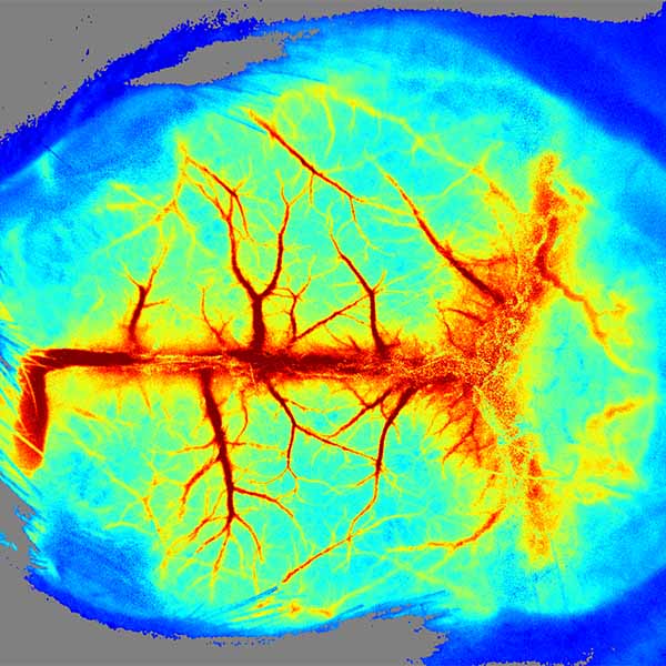
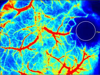
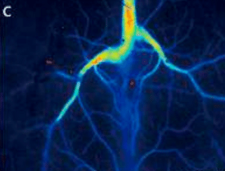
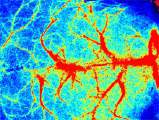
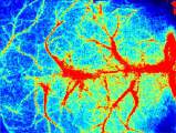
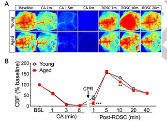
Detect the Changes of the Cerebral Cortex Blood Perfusion of a Cardiac Arrest (CA) and Return of Spontaneous Circulation (ROSC) Mouse Model
Detect the Changes of the Cerebral Cortex Blood Perfusion of a Cardiac Arrest
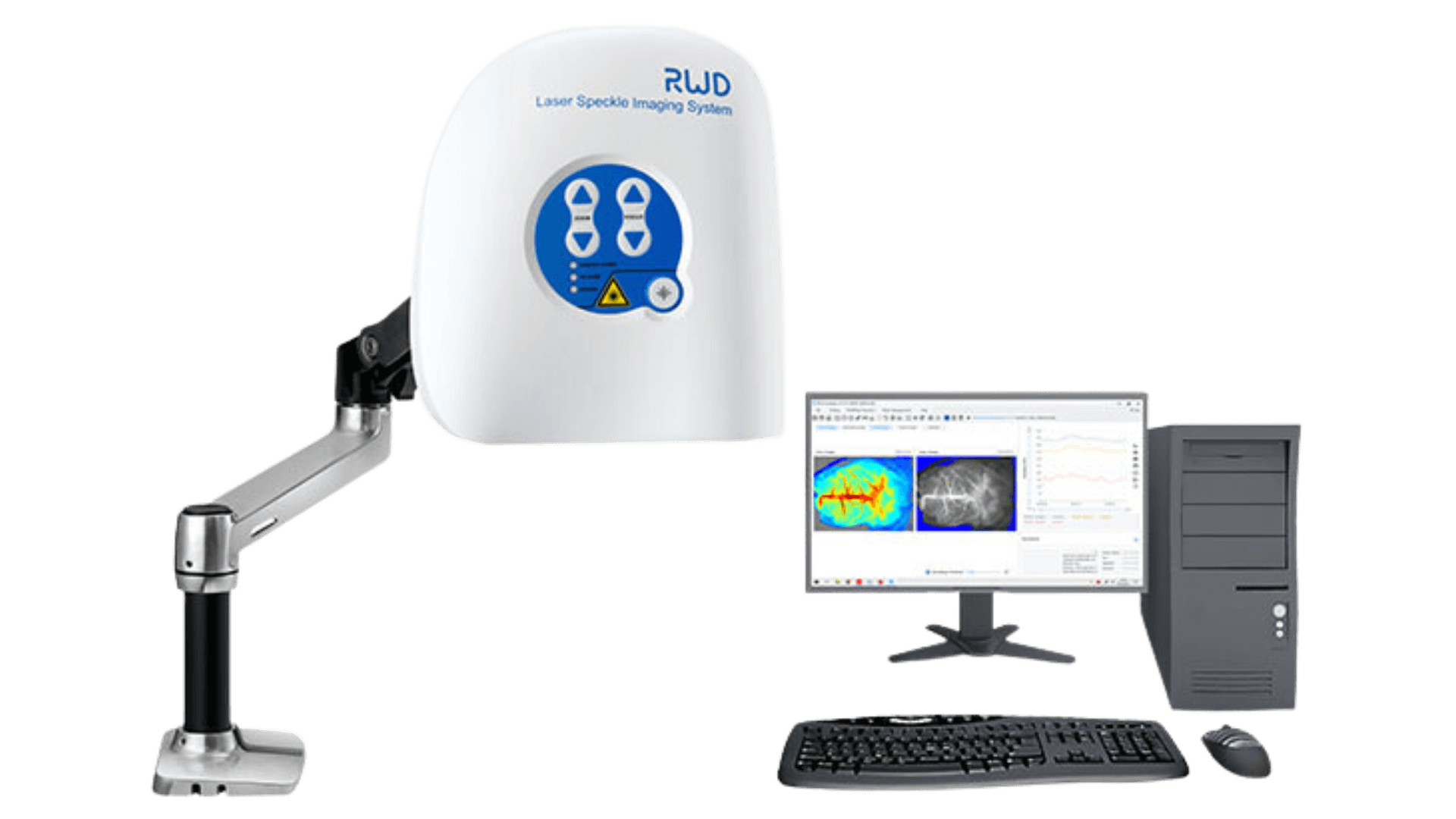
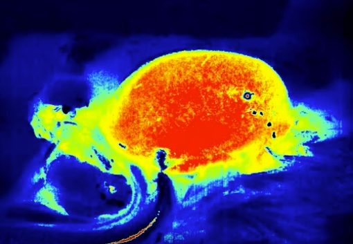
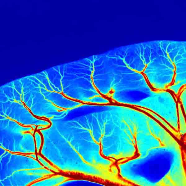
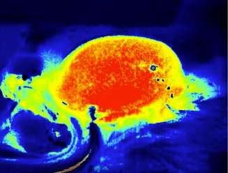
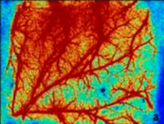
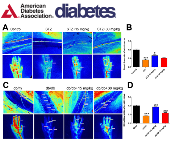
Real-Time Monitoring of the Regional Velocity, Distribution of Blood Flow, and Perfused Vessel for Sciatic Nerve and Foot Pads
Real-Time Monitoring of the Regional Velocity, Distribution of Blood Flow, Perfused Vessel for
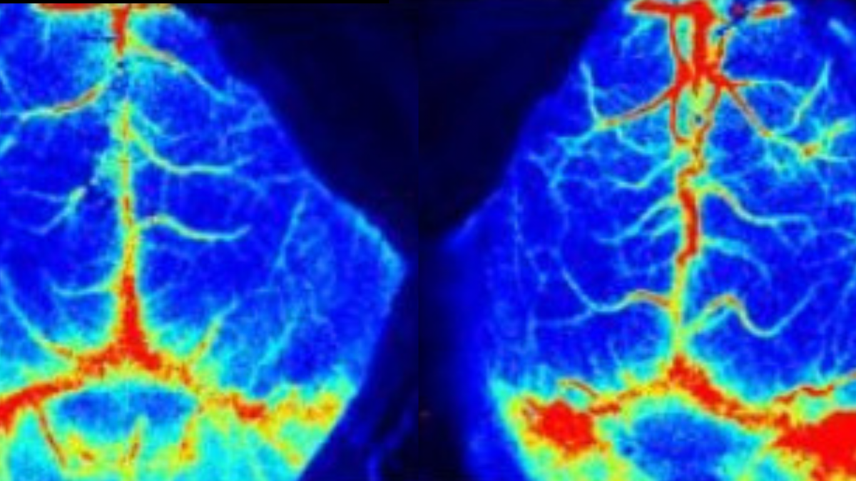
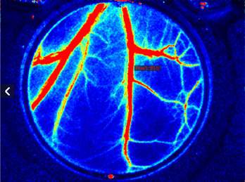
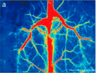
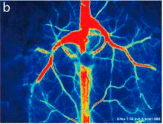
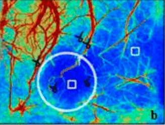
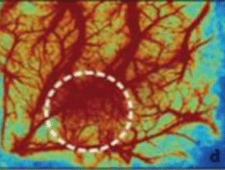
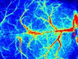
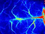
Using the Laser Speckle to Observe Changes in Cerebral Blood Flow after Lymphatic Ablation and Quantitative Analysis
Using the Laser Speckle Imaging System to Observe Changes in Cerebral Blood Flow