Article Review:
The Assessment of rhBMP-2 Loaded Bovine Hydroxyapatite Granules Using the SuperArgus Advance CT Systems by Sedecal
System Used:
SuperArgus PET/CT

Abstract
The purpose of this work was to validate the efficacy of a bone scaffold Proteo-Graft (Noricum S.L.). The granules of Proteo-Graft function for osteoconductive actions to support the adhesion and proliferation of mesenchymal stem cells (MSC). This scaffold’s recombinant human bone morphogenetic protein type 2 (rhBMP-2) component expediates osteoprogenitor differentiation. Noricum S.L. aims to study the efficacy of this bone scaffold with various biomaterials in vivo and the effect on critical bone lesions. Lopez-Andaluz, J and co-workers investigated the regeneration and adhesion to a biomaterial of this scaffold within the mandible bone of a rat with a critical bone defect.
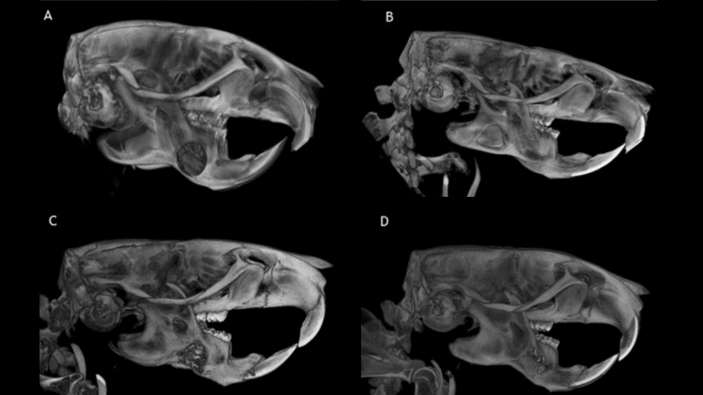
The SuperArgus Advance CT system was used to assess bone defects in vivo. The control group underwent all surgical procedures except no bone scaffold was used, just the biomaterial material in the defect area. The study group underwent surgery, and the lesion area was filled with the Proteo-Graft followed by being covered with the biomaterial. The bone defect was clearly visible within the control group at both 3- and 6-months post-surgery (figure 1). Within the study group there was no adhesion of the bone graft to the biomaterial within the first 3 months post-surgery. However, 6 months post-surgery the experimental material revealed enhanced osseointegration and there was difficulty seeing a difference between the defect and biomaterial (figure 2).
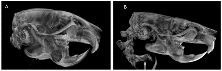
Figure 1. microCT images of the rat mandible in vivo. This control group consists of the biomaterial, with no bone scaffold for regeneration. A) This is a representative image 3-month post-surgery with no visible new bone formation (highlighted by a white arrow). B) This is a representative image 6-month post-surgery with visible new bone formation along the defect borders (highlighted by a white arrow).
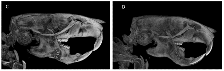
Figure 2. microCT images of the rat mandible in vivo. This is the study group consisting of the biomaterial and Proteo-Graft bone scaffold. A) This is a representative image 3-month post-surgery with visible new bone mineralization (highlighted by white arrow). B) This is a representative image 6-month post-surgery with visible new bone mineralization and clear adhesion to the biomaterial (highlighted by white arrow).
The SuperArgus CT system was able to visual the small defect area of 5 x 2 mm (diameter x depth) of the rat mandible near the alveolar zone in vivo. The CT clearly identify that at 3 and 6 months within the study group there was a noticeable breakdown of the biomaterial accompanied by new bone formation. The authors confirmed their findings via histological analysis post-mortem. They can confirm that this experimental scaffold promotes structural restitution of the critical bone defect. Further studies will be done to confirm therapeutic dosages and other variables that could play a role in bone regeneration.
References
- López-Andaluz, J., et al. “Assessment of rhBMP-2-loaded bovine hydroxyapatite granules in the guided bone regeneration of critical bone defect in rat mandible bone.” Journal of Dental Sciences (2023).
Author
Tyler Lalonde PhD, Product Manager at Scintica