How the Prospect T1 high-frequency ultrasound system can be used in preclinical developmental biology applications
System Used:
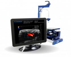
Prospect T1
This white paper will review the variety of ways the Prospect T1, high-frequency ultrasound system, can be used for preclinical applications in developmental biology non-invasively.
Specifically, this paper will review the following:
- Confirmation of Pregnancy
- Counting & Viability/Resorption of Embryos
- Embryo Staging
For those studying embryology, including embryo development, effect of transgenic mutations, and possible defects including embryonic lethal mutations, the ability to non-invasively visualize the embryos longitudinally in utero, along with the internal structures and organs as they develop, is instrumental. This brief white paper discusses some of these examples.
What is not mentioned here, but will be in future white papers, is the ability to use the same system to perform image guided needle injections into the externalized embryos to alter development in some capacity, and to then study the effects of this modulation over time. The embryos can be returned to the dam following the injection, to complete development and ultimately be born as pups. The development of the individual pups can then be followed over an appropriate time into adulthood.
1. Confirmation of Pregnancy
Precise embryonic timing is vital for many research applications (genetic engineering, tissue culturing, genetic and environmental factors influencing development, etc.). And many researchers have tried to find ways to confirm pregnancy at the earliest possible time while not reducing accuracy or increasing unnecessary invasiveness. If you take a quick scenario of breeding to achieve a specific sex of a double transgenic mouse, about one in every ten pups born will be the genotype and sex needed for your research goals. However, getting into other more complex and typical genetic schemes – animal waste can become even more extensive, especially if unnecessarily euthanizing dams that had a false pregnancy or using invasive means that may disrupt a current pregnancy. For that reason, researchers have sought ways to increase accuracy of pregnancy confirmation methods, but not necessarily at the expense of using more resources (i.e., study costs, employee time commitment, etc.); many of these methods are summarized below and in Figure 1.
Due to ease of results, the most commonly used confirmation method is to monitor for a vaginal plug, which is coagulated and hardened secretions from the male formed about 8-24 hours after breeding. Although this is a handy indication of sexual activity, it is not a definitive marker in determining a true pregnancy. In fact, using this method with inbred mice have false pregnancy rates that are reported to be up to 50% or higher1–3. Also, a vaginal plug is not always formed and can be easily dislodged. This method is simple and relatively easy to perform but can be entirely misleading.
With pregnancy, core body temperature increases, and changes in core body temperature measurements can detect pregnancy within 14 hours of initial pairing, even before embryo implantation. Unlike monitoring for a vaginal plug, core body temperature measurements can detect real pregnancy verse pregnancy-like events, giving it an accuracy rate of ~100%4. However, core body temperature measurements require the female breeders to have surgery before placing with a male to implant a small telemetry device to monitor these readings consistently, which is a time and financial commitment that isn’t feasible for most labs.
Throughout pregnancy, progesterone concentrations are known to increase steadily until shifting nearer to birth. Significant changes in Progesterone concentration measurements are early (about day 2-4)5. However, this measurement comes at the expense of being invasive, although minimally. Progesterone is transported in the body entirely by plasma, so blood needs to be drawn, and depending on the protocol, administration of anesthetics becomes necessary, all of which run the risk of disrupting the pregnancy.
Ultrasound is a reliable and non-invasive means to confirm pregnancy as early as 4.5 to 6.5 days, depending on the system’s frequency and resolution capacity. With high-frequency ultrasounds, an embryo/fetus (hereafter referred to as an embryo for simplicity) is quickly noticeable to confirm pregnancy, can be counted for an estimation of litter sizes, can be imaged to precisely estimate age or gestational stage, can show the viability of these embryos as well as the morphology and functioning of many organs. The short duration of ultrasound examination makes it a quick process and also avoids the risk of inducing changes in pre-and post-natal growth and development6. However, proper orientation or position of the ultrasound probe is needed for accurate and consistent measurements. Overall, this method allows for precise confirmation of pregnancy early, accurately, and easily.
Changes in the dam’s physical characteristics are easily noticeable by a trained technician. Within 7.5 days, pregnant females will gain 3x as much bodyweight as compared to nonpregnant females3. Weight gain is a relatively easy and non-invasive means of confirming pregnancy, but with reduced accuracy of detecting a true pregnancy as well as detecting the true gestational age of the embryos. Also, at about 14 days, the pups can physically be palpated in the abdomen, confirming true pregnancy. However, for many researchers, both timepoints for confirming pregnancy are too late to be helpful markers within their work.

Figure 1: Different Methods for Confirming Pregnancy. Illustrated are currently known methods for confirming pregnancy and the earliest date each of these methods can be performed. *The mouse is currently the gold-standard research model, and the gestation period of a typical mouse embryo is modeled here on this timeline; technology generalities can be applied to most species, but specific timeline differences will be noticeable. This timeline is not drawn to scale. GD= gestational day.
2. Counting & Viability/Resorption of Embryos
Ultrasound systems can count and track specific positions of embryos in utero over multiple days if desired. A researcher can identify any set of embryos, normal or abnormal, and see how each embryo changes based on their research variables. For example, researchers used Listeria monocytogenes – known to cause a toxic infection in infected pups, causing reabsorption by the female dam. This is a common induced-miscarriage mouse model. Using ultrasound, the researchers showed that an abnormally slow heart rate was induced in the infected pups, and that the dam had reabsorbed the pups before birth of the others7.
Although counting and specific positioning of embryos may be necessary for certain research goals, it is important to keep in mind that embryos, especially in mice, tend to wind and contour themselves into unique locations within the dam. After gestation day (GD) 10, ultrasound imaging of embryos can be a challenge for even someone who is quite experienced with the technology, because the embryos will be sprawled around and at different depths in the dam’s abdomen. Another point that should be stressed is that high-frequency ultrasound systems (equipped with around 20-40 MHz probes) should be used for this procedure so that these small embryos can be visualized clearly, and any excess ambiguity is removed. Consistency by trained technicians should also be stressed since it can be easy to miss an embryo if not searched correctly and getting exact counts of embryos can be quite challenging due to underestimation8. Overall, high-frequency ultrasound systems can be a quick, easy, and reliable method to count and/or track positioning of embryos in utero.
3. Embryo Staging
Ultrasound is also useful in determining the gestational age of the embryo. Embryonic development happens on a well-defined timeline for each species with slight variations due to circumstances. A mouse embryo’s timeline, for example, can vary based on the different rates of development between mouse strains, other environmental conditions applied, like lighting, diet, etc.9. Knowing your specific species gestational timeline is vital to many studies and ultrasound has made figuring out those timelines possible. In mice, for example, using ultrasound and taking a simple length measurement from crown to rump of the pups, the precise gestational age can be estimated with fairly high accuracy using a linear regression model, where each increase in 1 mm in crown-rump length correlates to an increase in age of 0.5d10. Others have shown similar results in the decidual sac size, gestational sac length, gestational sac thickness, placental length, etc. correlating well with gestational age11,12. However, many researchers will visualize the embryo and be able to quickly estimate what stage it is in without using fine-tuned measurements. Either way, ultrasound is a quick and effective way to visualize or confirm the gestational stage that the embryos are in, which can be vital to many timed-pregnancy studies.
Pre-Implantation Period
At this stage (GD0-3), the fertilized one-cell egg, divides into the beginning stages of a multiple-cell embryo and will implant within the uterine wall (GD0-4.5) Ultrasound is not able to visualize this process or embryos within this gestational stage to the degree needed to get quality data; however, after this stage depending on the frequency and resolution of the ultrasound system, the embryo can be easily and clearly imaged.
Post-Implantation Period
Correct development in utero happens in the post-implantation period and is critical to the survival and the overall health of the organism throughout its’ lifespan. Abnormal development has caused many mouse models to suffer from early embryonic lethality-ultimately halting research on that specific model. Ultrasound allows for the unique ability for researchers to study the morphological and functional capacity of specific organs, which helps to give clues as to the viability of specific transgenic animals, cause of congenital defects, or effects of physiological conditions on prenatal development, etc.
Morphological and anatomical landmarks that can be imaged using ultrasound will be summarized within this paper – a few of these landmarks are shown using ultrasound images in Figure 2; however, for an in-depth overview of the staging of mouse embryos, morphological and anatomical landmarks that can be imaged, etc., please refer to The Mouse Embryonic Imaging Guide available on the Scintica website (https://www.scintica.com/wp-content/uploads/2019/02/The-Mouse-Embryonic-Imaging-Guide_20140721_final.pdf).
In brief, researchers can confirm pregnancy around GD4.5-6.5, because a cylindrical embryo is formed. By GD7.5, the embryo has developed three distinct cavities: the amniotic, the exocoelomic, and the ectoplacental cavity. Starting at GD8.5, the morphogenesis and functioning of principal organs, such as the heart, kidney, liver, brain, eyes, etc., can be examined. The heart is the first organ to develop at GD8.5, and early cardiac measures, like cardiac inflow and outflow waveforms, can be taken around GD9.5 and from GD11-14 will resemble normal human fetal cardiac development at 7-20 weeks gestation13–16. The brain starts to develop at GD8.5, and early morphological structures can be imaged. Also, direct manipulation of mouse embryos has been difficult because the embryos are encased in the maternal uterus, but ultrasound–guided embryonic injections easily allow for a less invasive means of inducing specific transgenic animals. At about GD10.5-11.5, blood flow in the umbilical cord, the placenta, the ascending aorta, descending aorta, and dorsal aorta can be imaged. Researchers have started a comprehensive database for echocardiographic and Doppler parameters of embryonic mouse cardiovascular function from 12.5 to term15. Databases like these can serve as a standard for evaluating cardiac development and blood flow patterns in genetically altered mouse models15,16 Preclinical, high-frequency ultrasound systems are needed to get these early measurements; clinical ultrasound systems have reduced 2D spatial resolution and can only be reliably used in later stages of mouse embryonic development15.
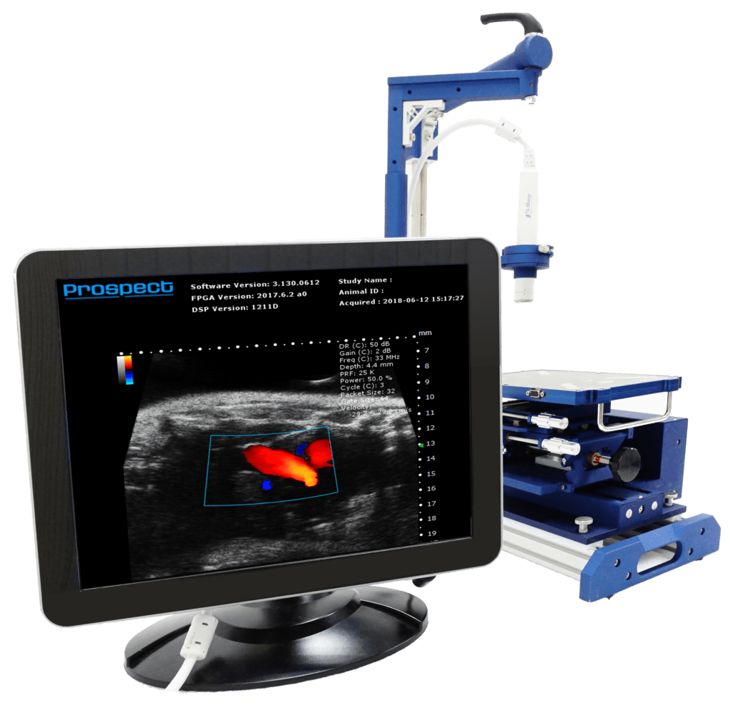
We provide a high-frequency (20-50MHz) and high resolution (~30μm) ultrasound imaging system called the Prospect T1. The Prospect T1 is capable of applications cited throughout and we always welcome questions about any specific research and/or applications. Check out the Prospect T1 High-Frequency Ultrasound System website page or reach out to Scintica Instrumentation if you would like to discuss your specific research needs (https://www.scintica.com/products/s-sharp-prospect-t1/).
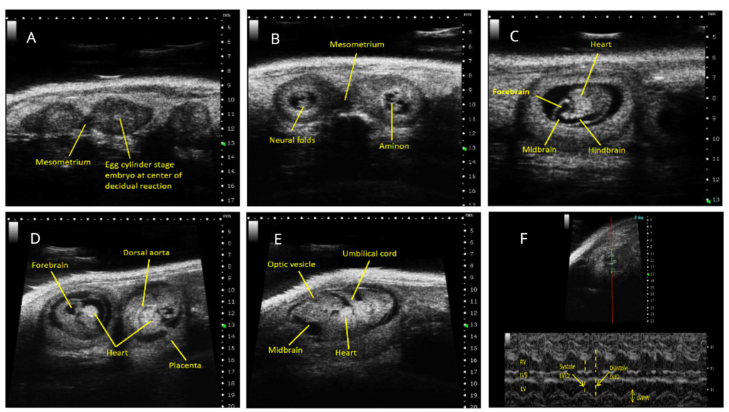
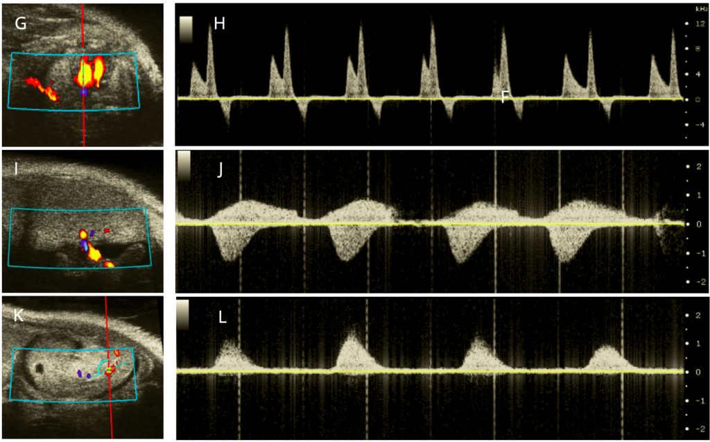
Figure 2: Ultrasound Images across embryonic development in utero. Ultrasound B-Mode images illustrating A) confirmation of pregnancy and counting of pups (3 pups shown; GD6.5), B) open neural folds (2 pups; GD8.5), C) heart, forebrain, midbrain, and hindbrain (1 pup; GD9.5) D) heart, forebrain, dorsal aorta, and placenta (2 pups; GD10.5), and E) heart, midbrain, optic vesicle, and umbilical cord (1 pup; GD11.5). Ultrasound M-Mode images illustrating F) cardiac measurements (1 pup; GD16.5). Ultrasound doppler-mode imaging of the G) apical four-chamber view, the pulse wave doppler image of H) blood flow through the Mitral valve, a reliable parameter for diastolic cardiac functioning, the I) view of the umbilical cord, J) Umbilical cord blood flow and K) view of the Dorsal aorta, and the L) Dorsal aorta blood flow. GD= gestational day.
References
- Biology of the Laboratory Mouse. http://www.informatics.jax.org/greenbook/.
- Lee Silver’s Mouse Genetics. http://www.informatics.jax.org/silver/.
- Heyne, G. W. et al. A Simple and Reliable Method for Early Pregnancy Detection in Inbred Mice. Journal of the American Association for Laboratory Animal Science vol. 54 (2015).
- Smarr, B. L., Zucker, I. & Kriegsfeld, L. J. Detection of Successful and Unsuccessful Pregnancies in Mice within Hours of Pairing through Frequency Analysis of High Temporal Resolution Core Body Temperature Data. (2016) doi:10.1371/journal.pone.0160127.
- Piekorz, R. P., Gingras, S., Hoffmeyer, A., Ihle, J. N. & Weinstein, Y. Regulation of Progesterone Levels during Pregnancy and Parturition by Signal Transducer and Activator of Transcription 5 and 20α-Hydroxysteroid Dehydrogenase. Molecular Endocrinology 19, 431–440 (2005).
- Pallares, P. & Gonzalez-Bulnes, A. Non-invasive ultrasonographic characterization of phenotypic changes during embryo development in non-anesthetized mice of different genotypes. Theriogenology 70, 44–52 (2008).
- Hardy, J. et al. Infection of pregnant mice with Listeria monocytogenes induces fetal bradycardia. Pediatric Research 2012 71:5 71, 539–545 (2012).
- Pallares, P. & Gonzalez-Bulnes, A. Use of ultrasound imaging for early diagnosis of pregnancy and determination of litter size in the mouse. Lab Anim 43, 91–95 (2009).
- Staging of gastrulating mouse embryos by morphological landmarks in the dissecting microscope.
- Brown, S. D. et al. Comparative Medicine Ultrasound Diagnosis of Mouse Pregnancy and Gestational Staging 405206-3. (2006).
- Chang, C. P., Chen, L. & Crabtree, G. R. Sonographic staging of the developmental status of mouse embryos in utero. Genesis 36, 7–11 (2003).
- Greco, A. et al. High Frequency Ultrasound for In Vivo Pregnancy Diagnosis and Staging of Placental and Fetal Development in Mice. PLOS ONE 8, e77205 (2013).
- Srinivasan, S. et al. Noninvasive, in utero imaging of mouse embryonic heart development with 40-MHz echocardiography. Circulation 98, 912–918 (1998).
- Turnbull, D. H. In utero ultrasound backscatter microscopy of early stage mouse embryos. Comput Med Imaging Graph 23, 25–31 (1999).
- Yu, Q., Leatherbury, L., Tian, X. & Lo, C. W. Cardiovascular assessment of fetal mice by in utero echocardiography. Ultrasound Med Biol 34, 741–752 (2008).
- Gui, Y. H., Linask, K. K., Khowsathit, P. & Huhta, J. C. Doppler Echocardiography of Normal and Abnormal Embryonic Mouse Heart. Pediatric Research 1996 40:4 40, 633–642 (1996).
- Phoon, C. K. L. & Turnbull, D. H. Ultrasound biomicroscopy-Doppler in mouse cardiovascular development 1. (2003) doi:10.1152/physiolgenomics.00008.2003.-The.
- Nieto, M. et al. Antisense miR-7 impairs insulin expression in developing pancreas and in cultured pancreatic buds. Cell Transplantation 21, 1761–1774 (2012).
- Rinaldi, S. F. et al. Ultrasound-guided intrauterine injection of lipopolysaccharide as a novel model of preterm birth in the mouse. Am J Pathol 185, 1201–1206 (2015).
Prospect T1: High-Frequency Ultrasound