


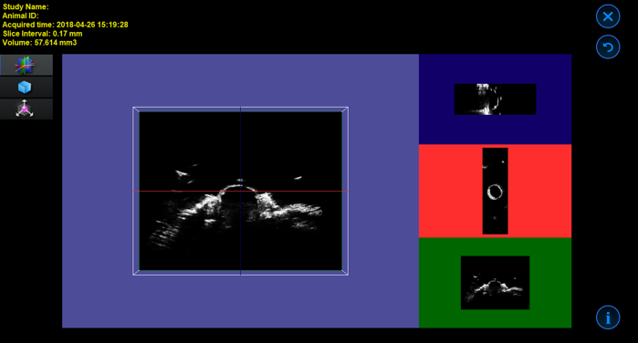
3D Motor and Acquisition/Analysis Software
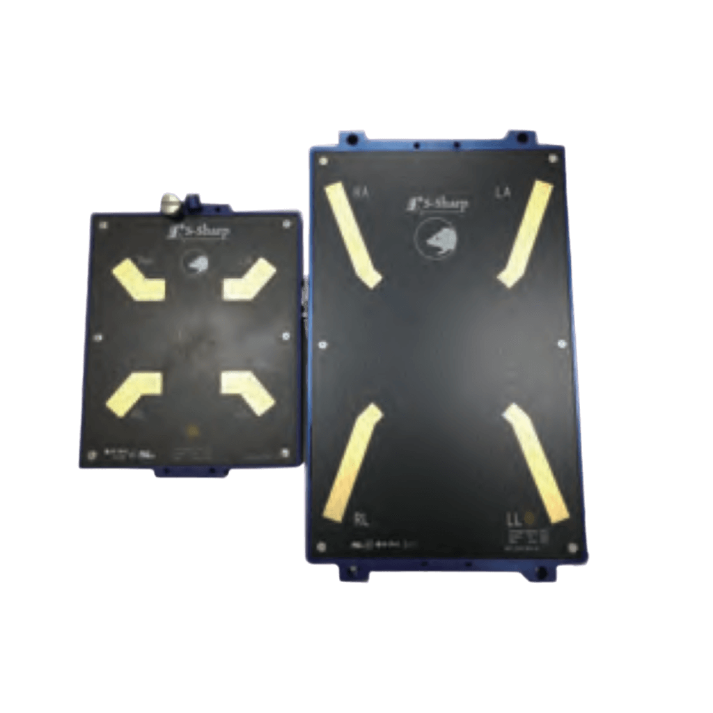
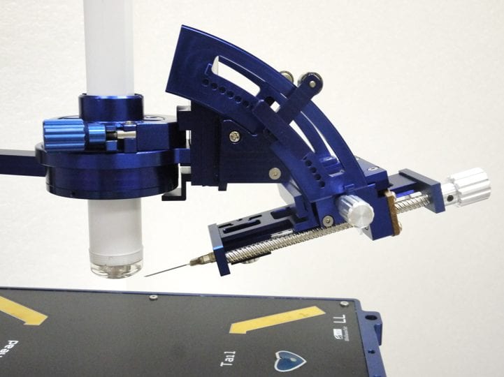
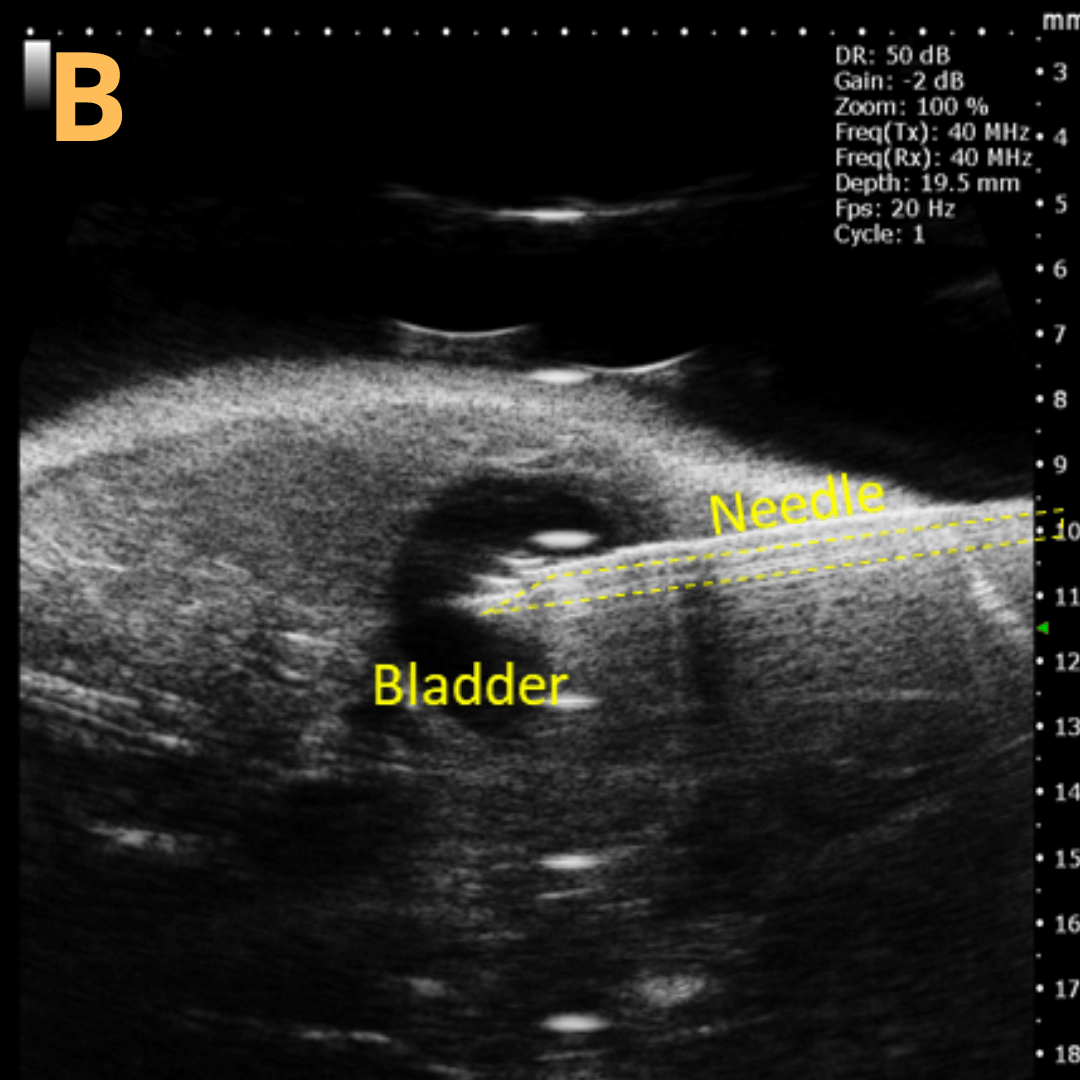
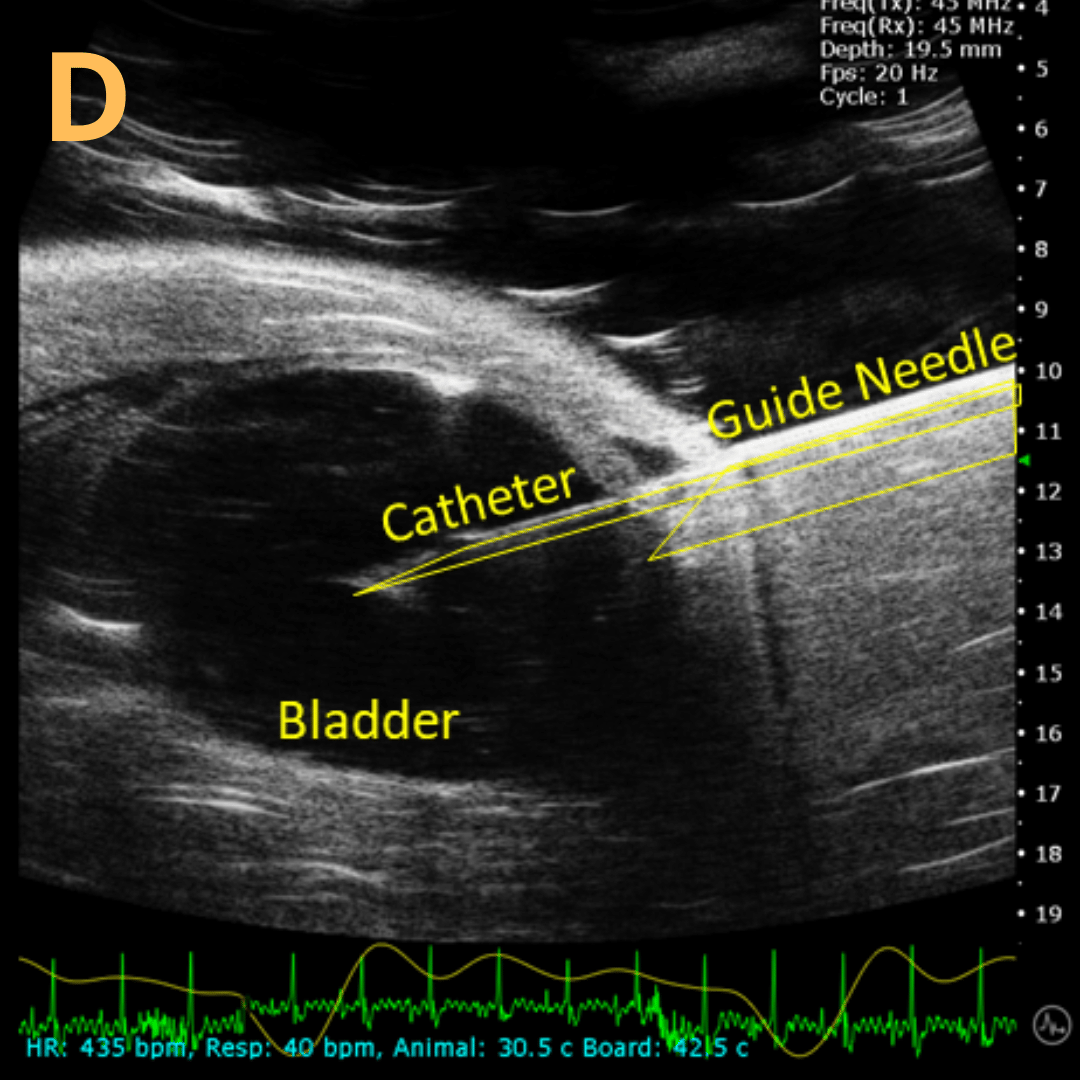
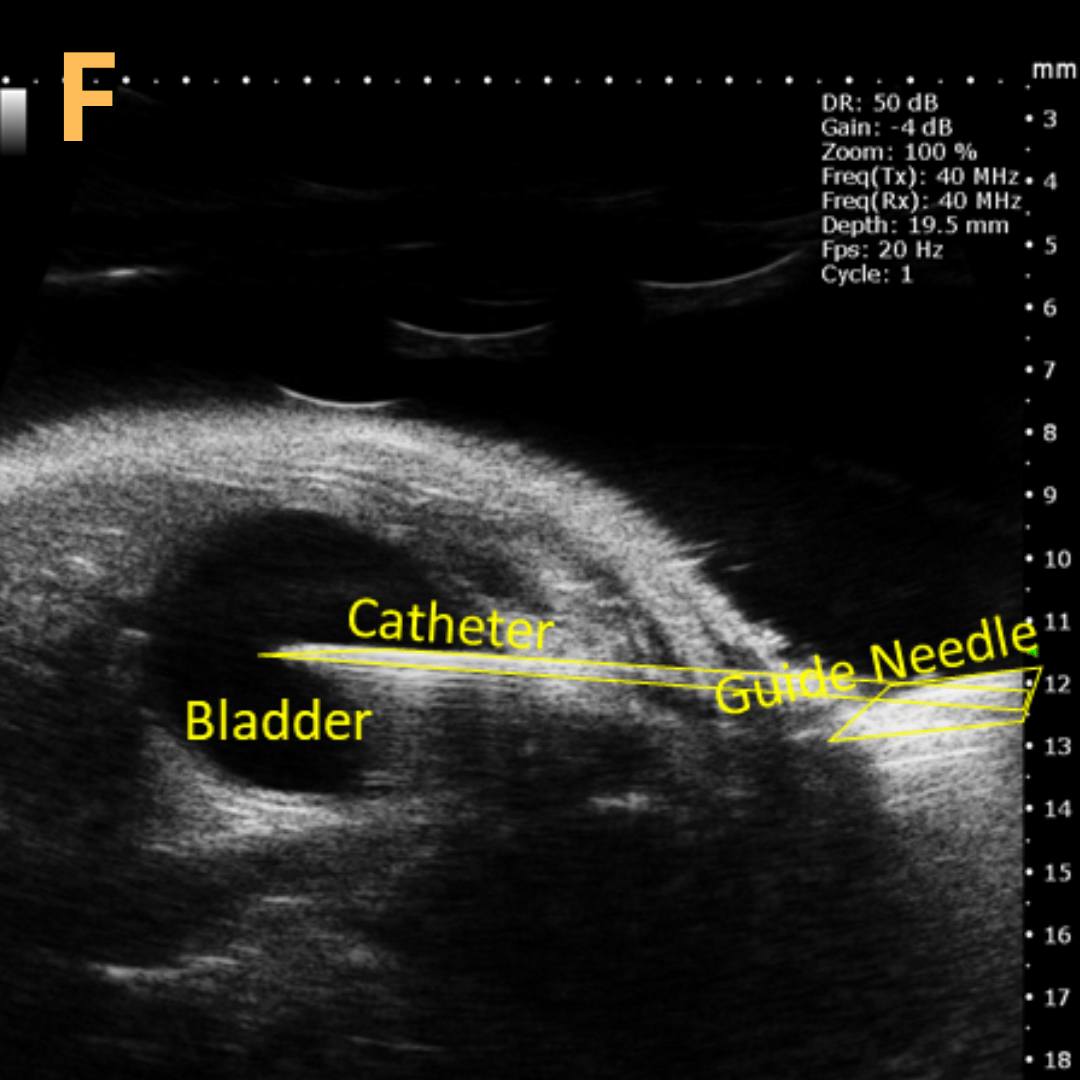
Image-guided Needle Injection Mount
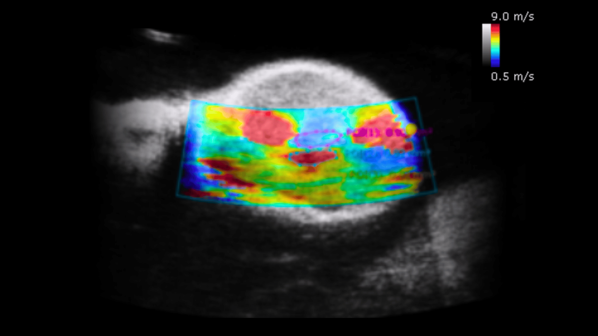
Integrated Shear Wave Elastography Probe and Analysis Software
3D Motor and Acquisition/Analysis Software
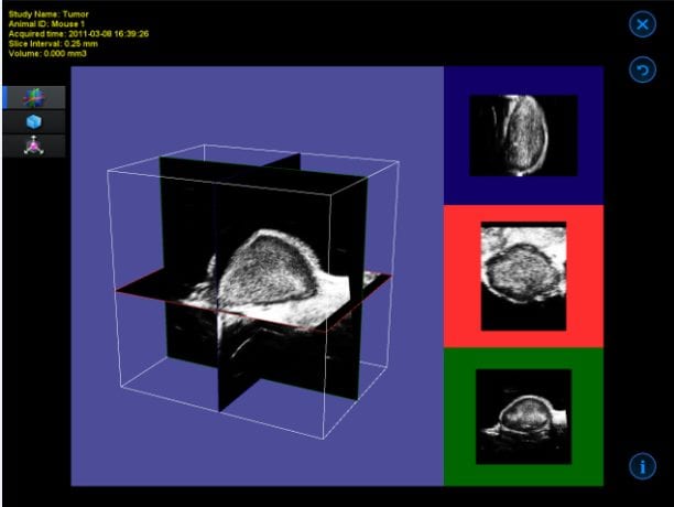
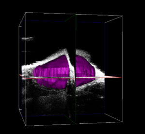
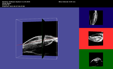
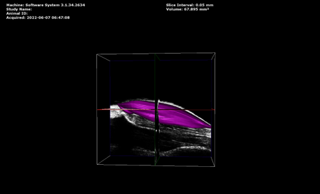
Image-guided Needle Injection Mount
Integrated Shear Wave Elastography Probe and Analysis Software
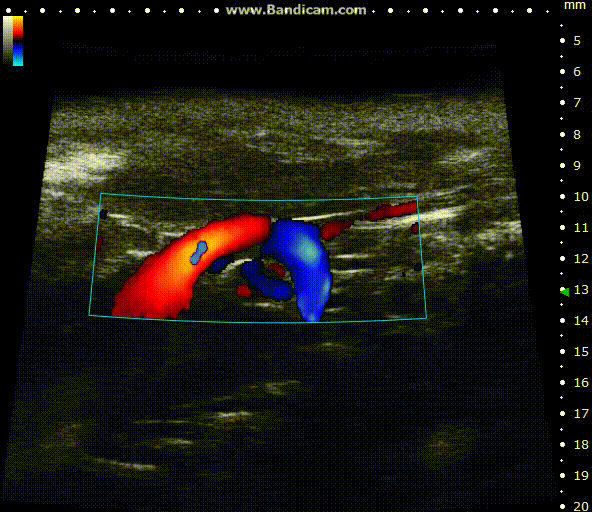
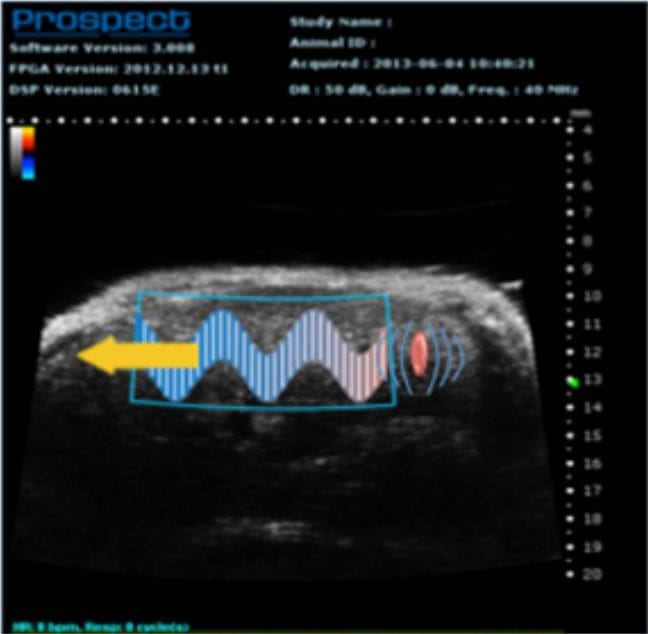
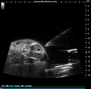
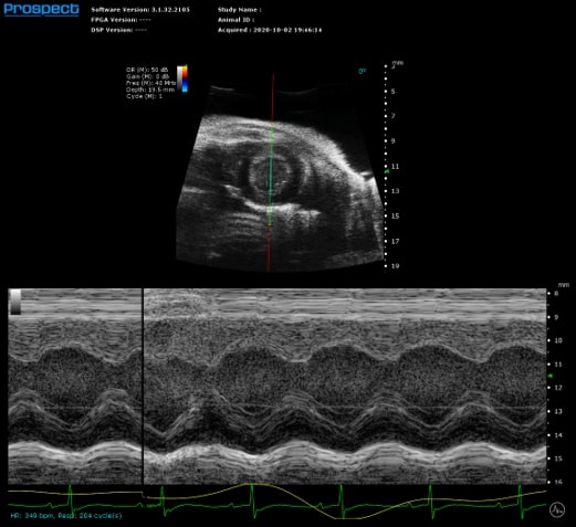
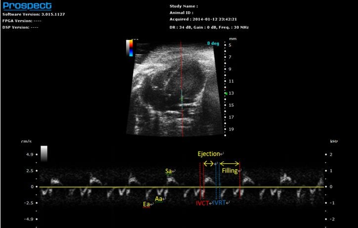
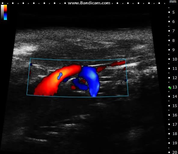
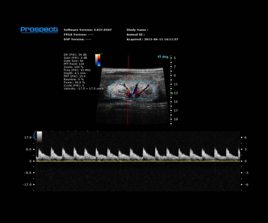
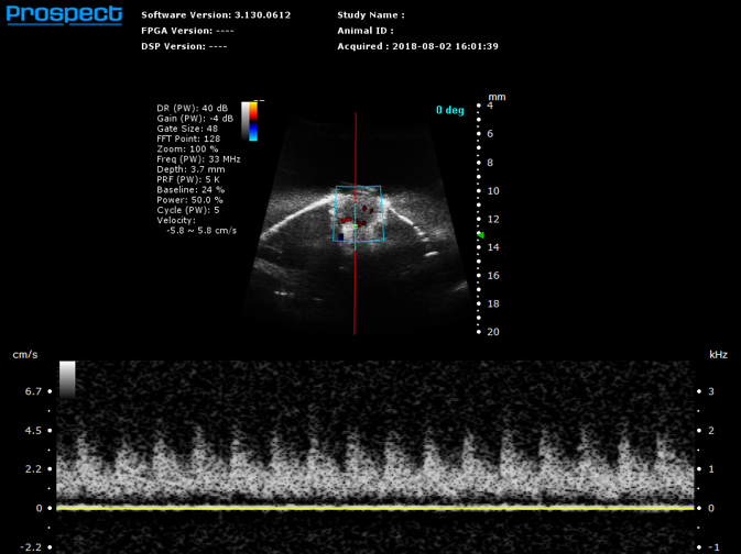
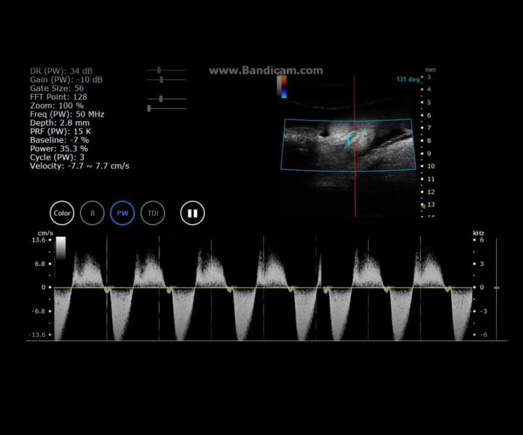
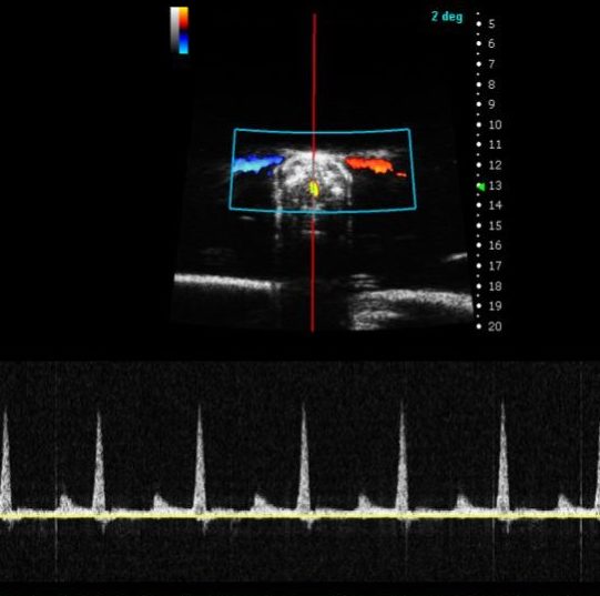
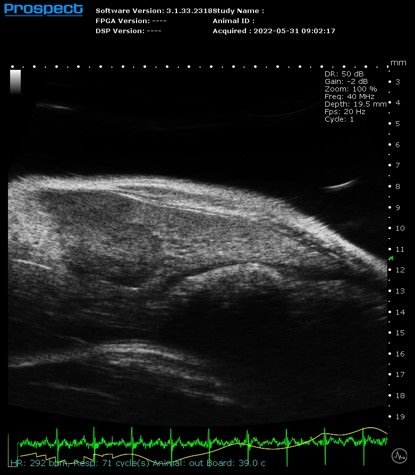
Cardiovascular Biology
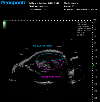
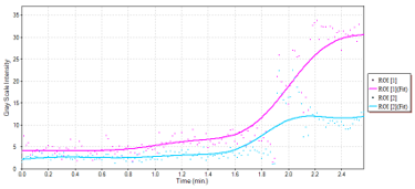
Cancer Biology
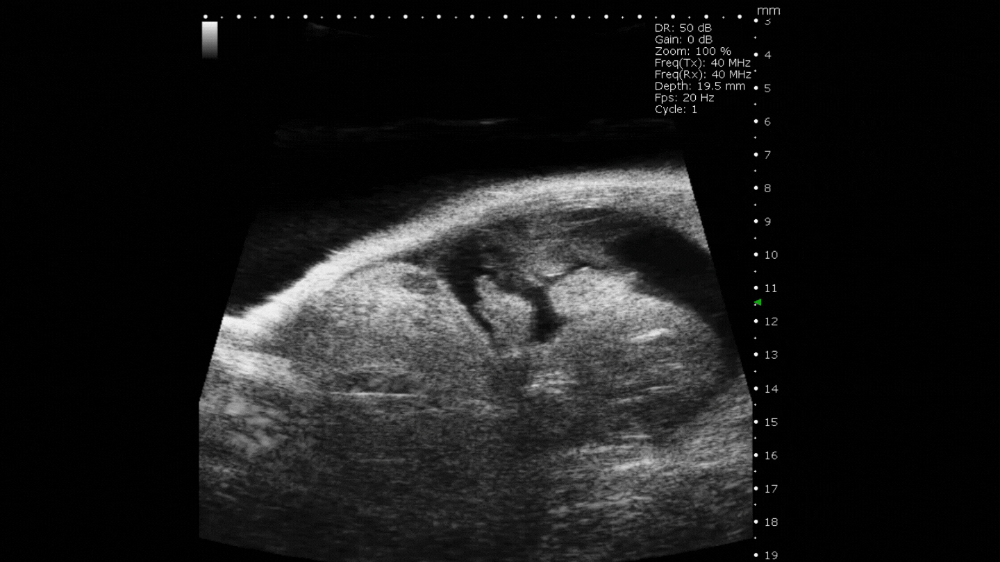
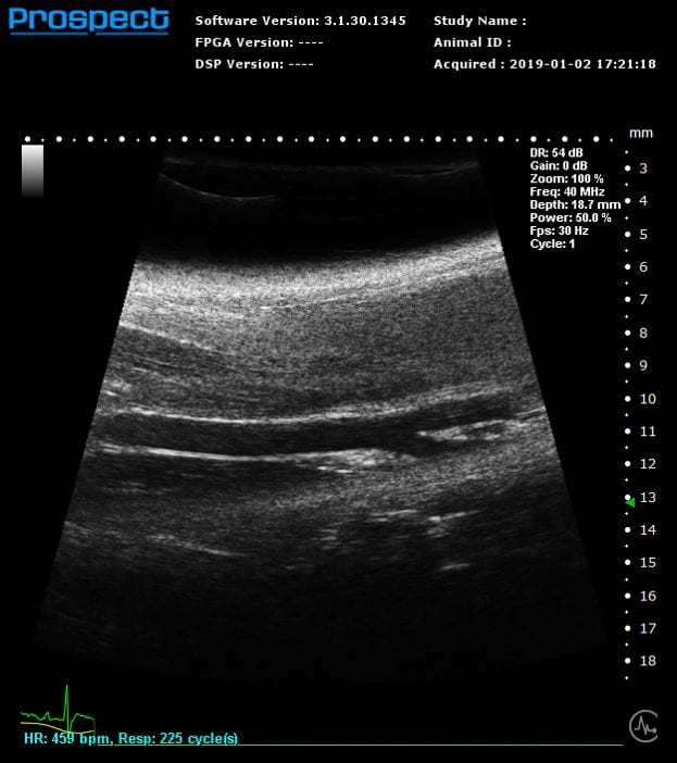
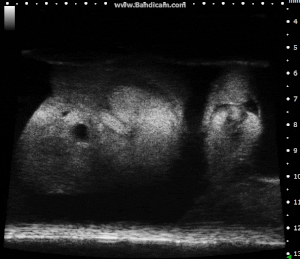
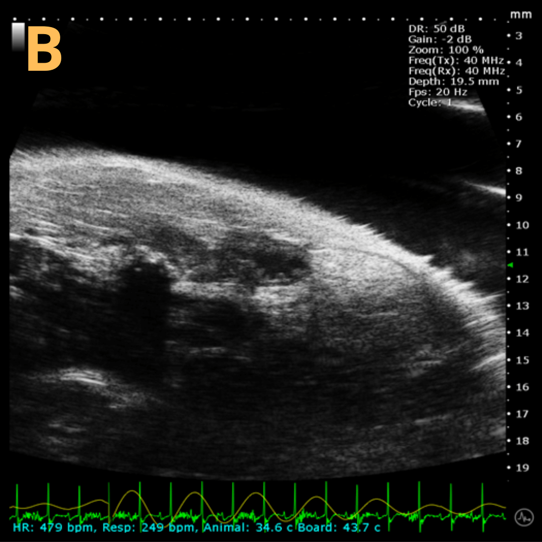
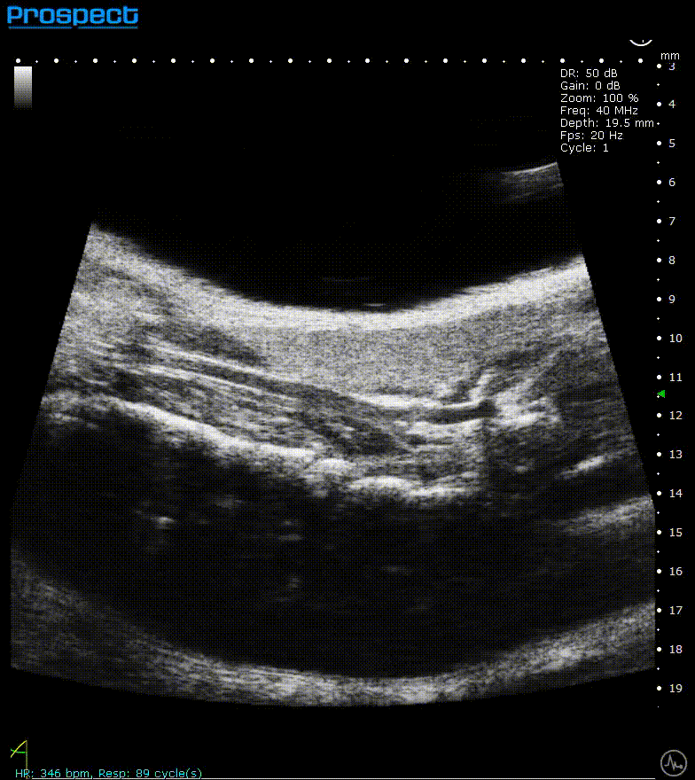
Abdominal & Anatomical Imaging
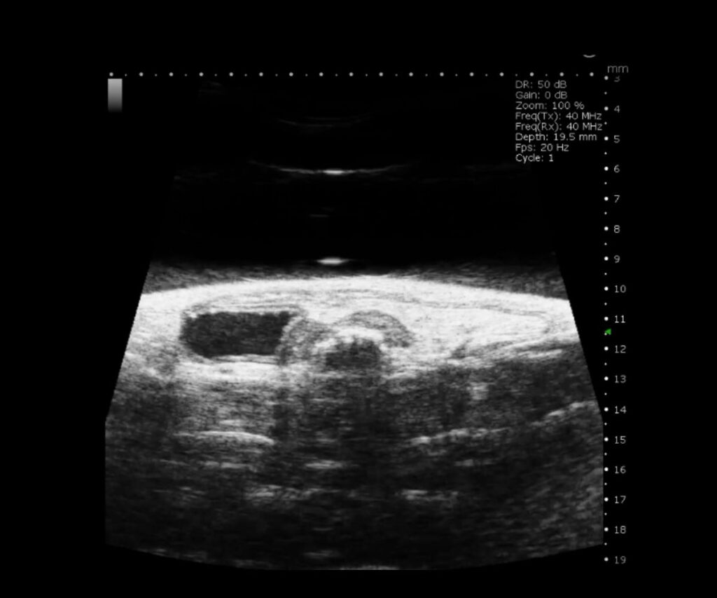
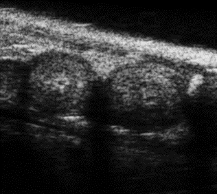
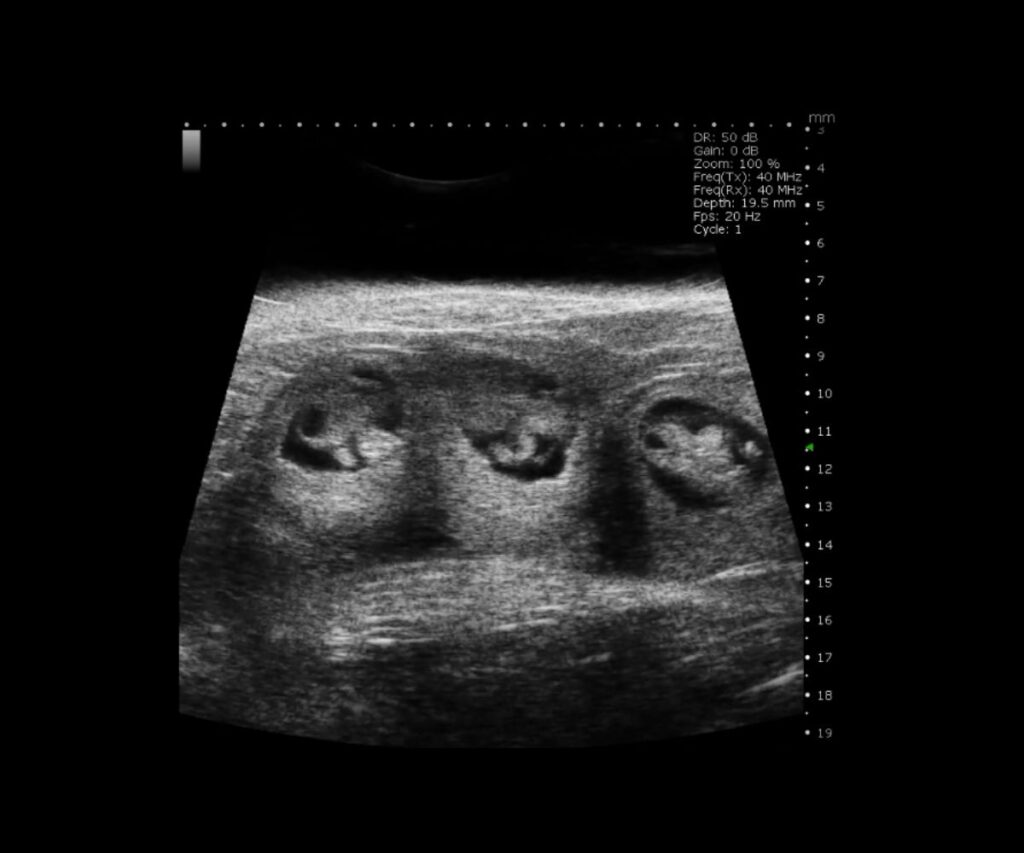
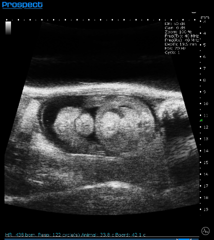
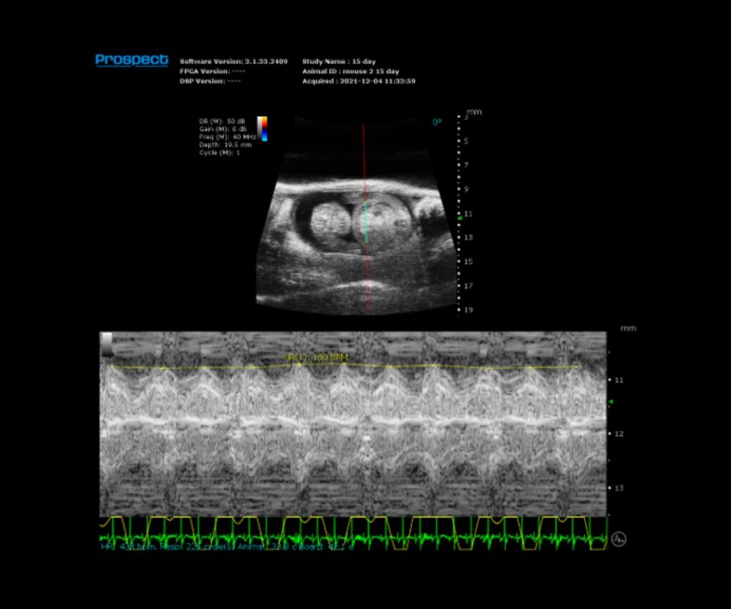
Developmental Biology
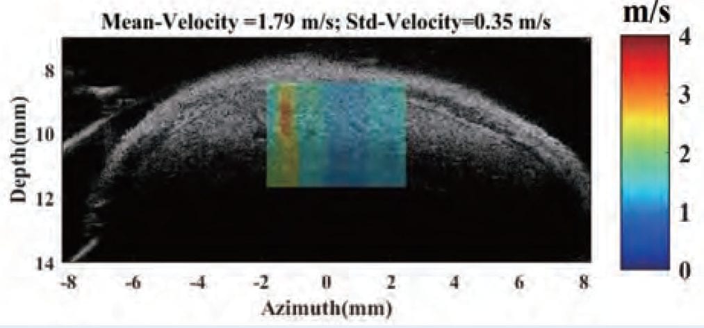
Ophthalmology
Other Species
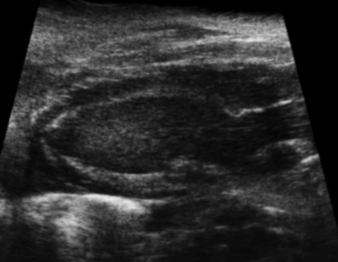
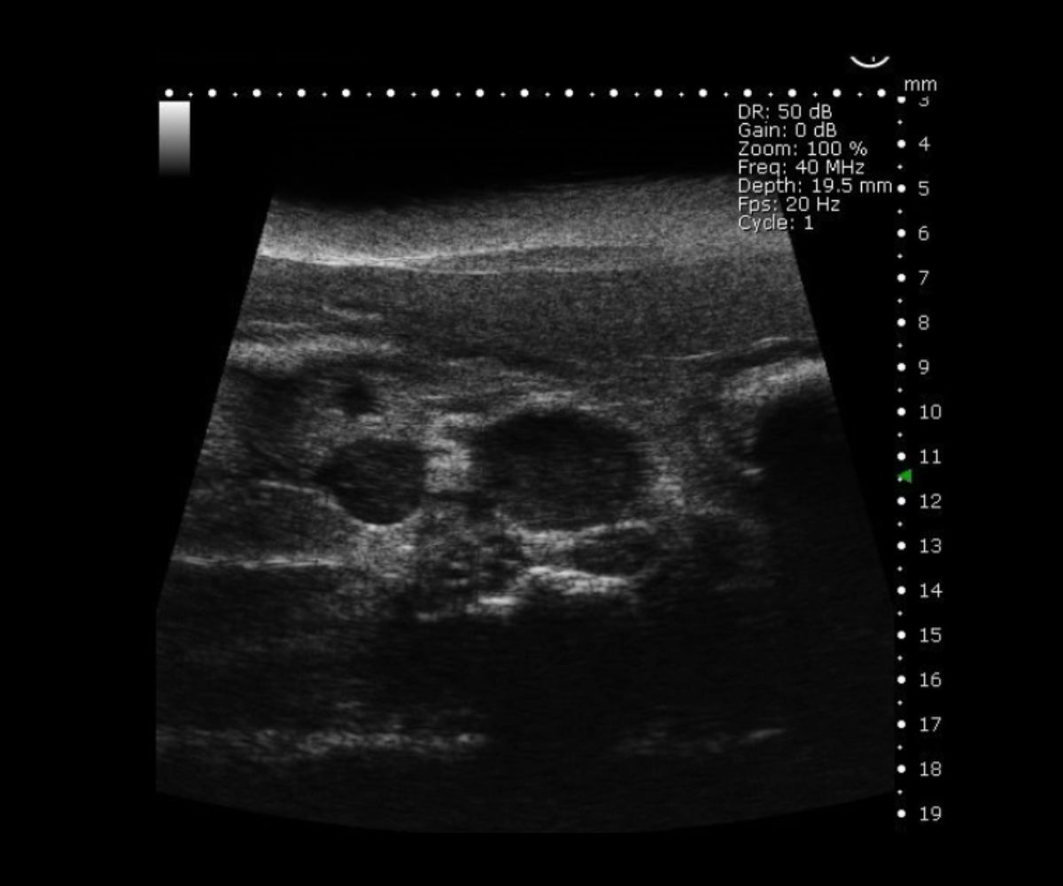
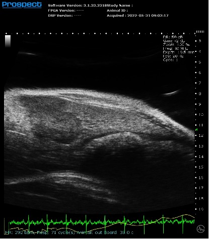
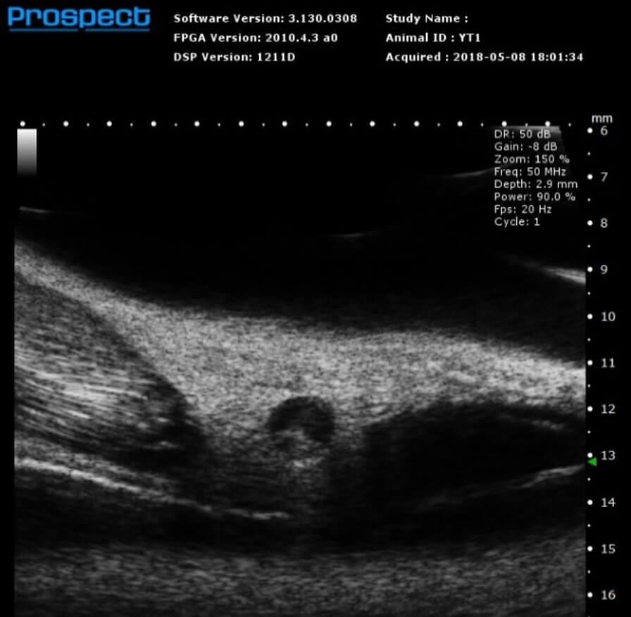
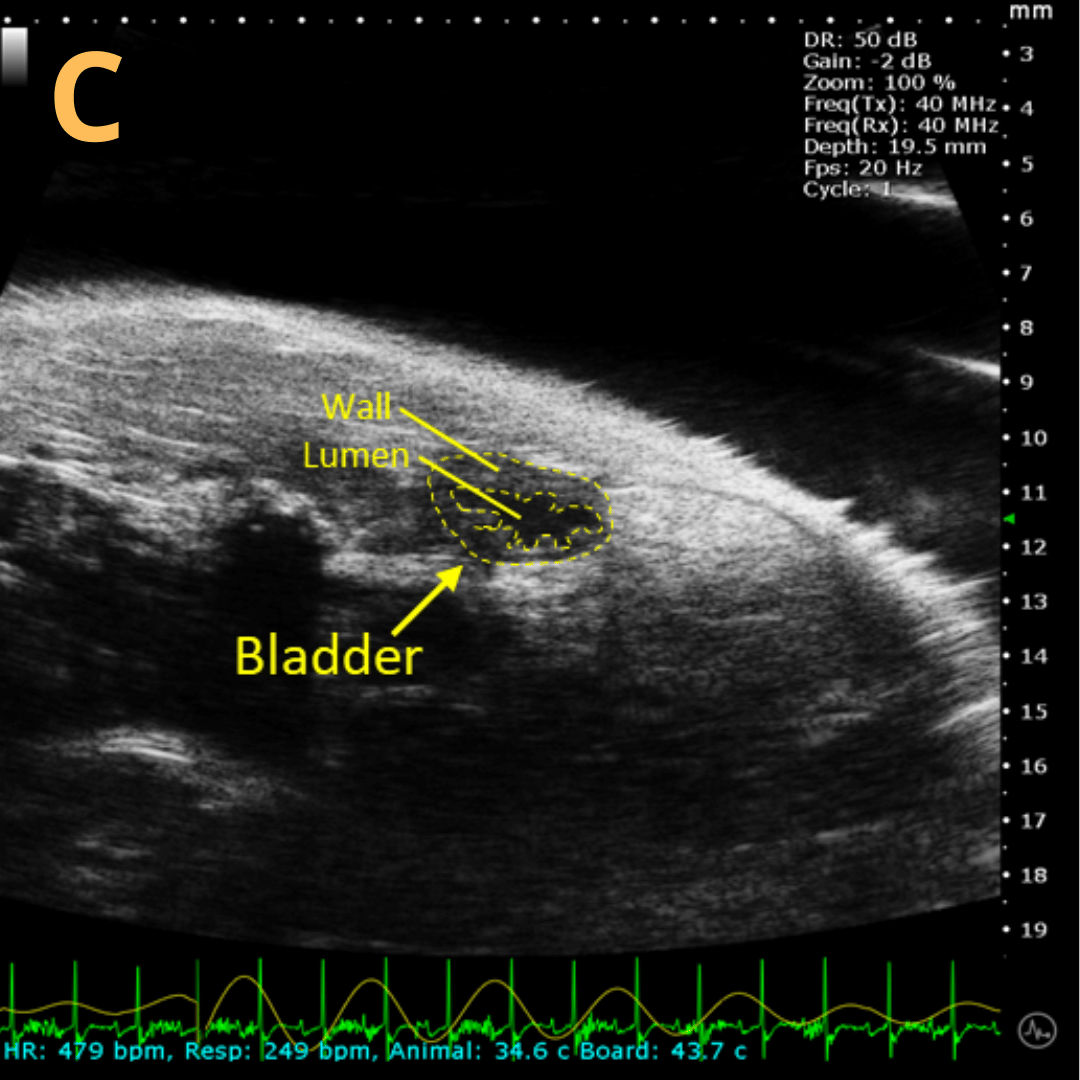
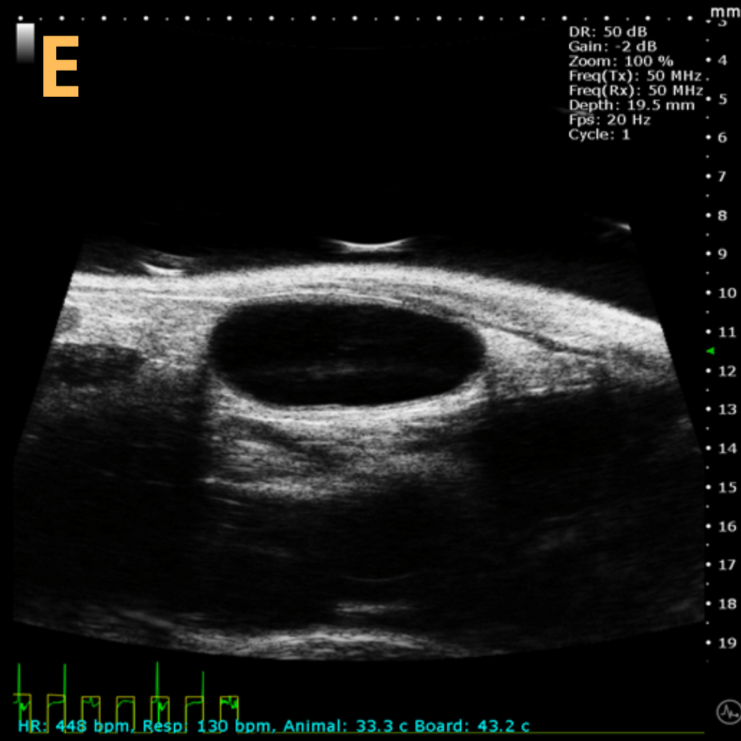
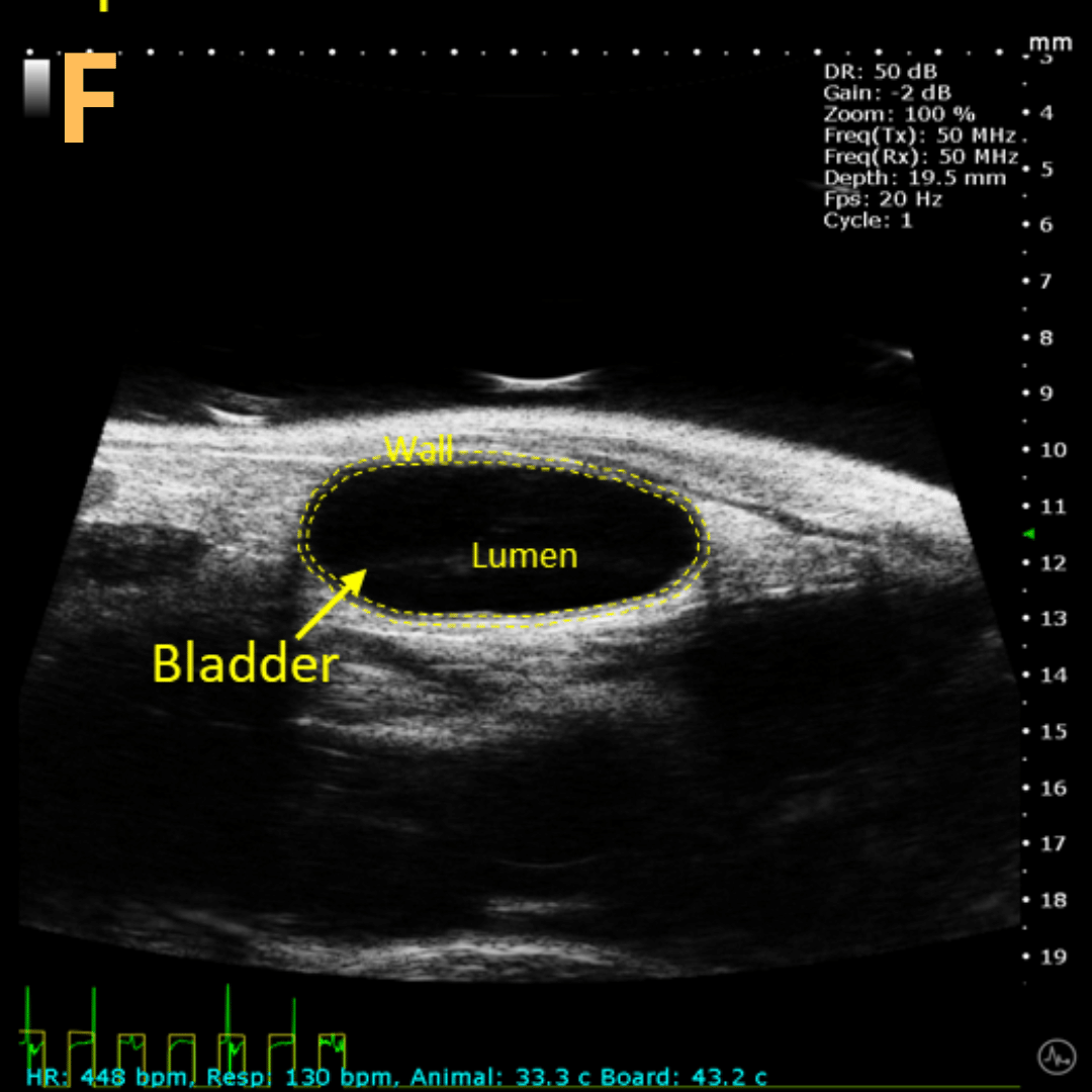
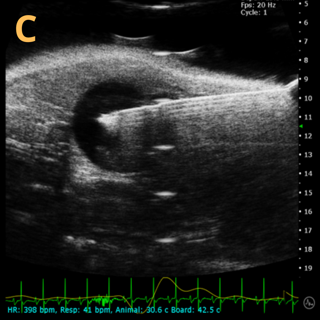
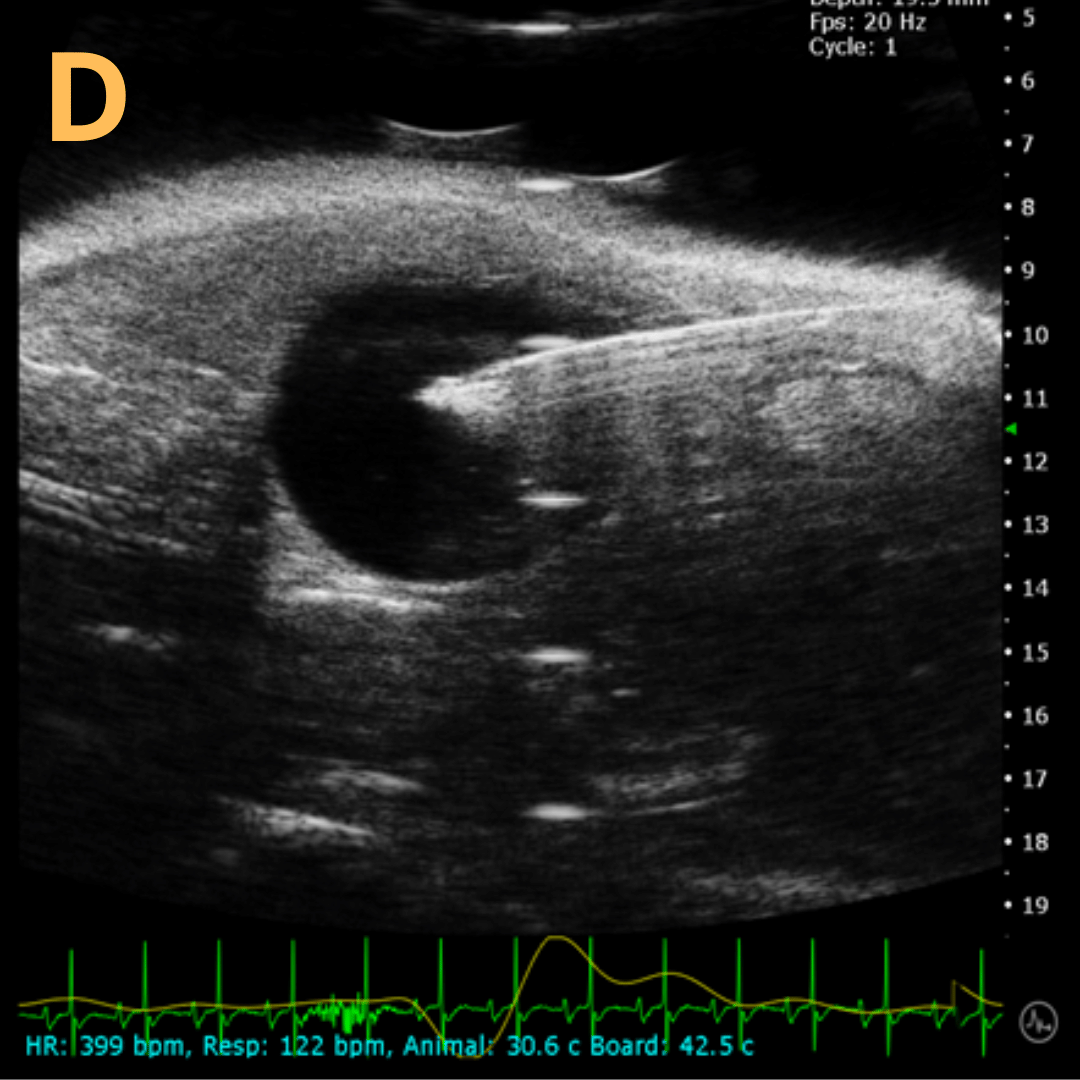
Bladder Imaging
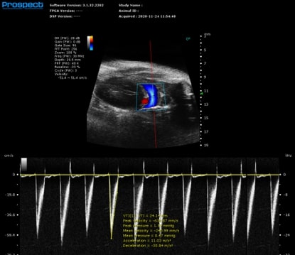
Cardiovascular Biology


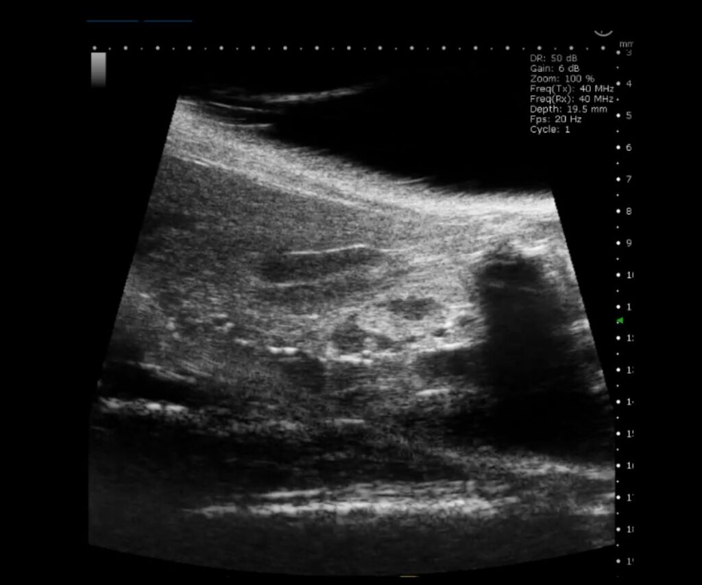
Cancer Biology
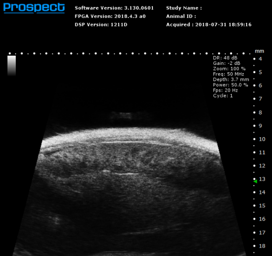
Abdominal & Anatomical Imaging

Developmental Biology
Ophthalmology
Other Species
Bladder Imaging
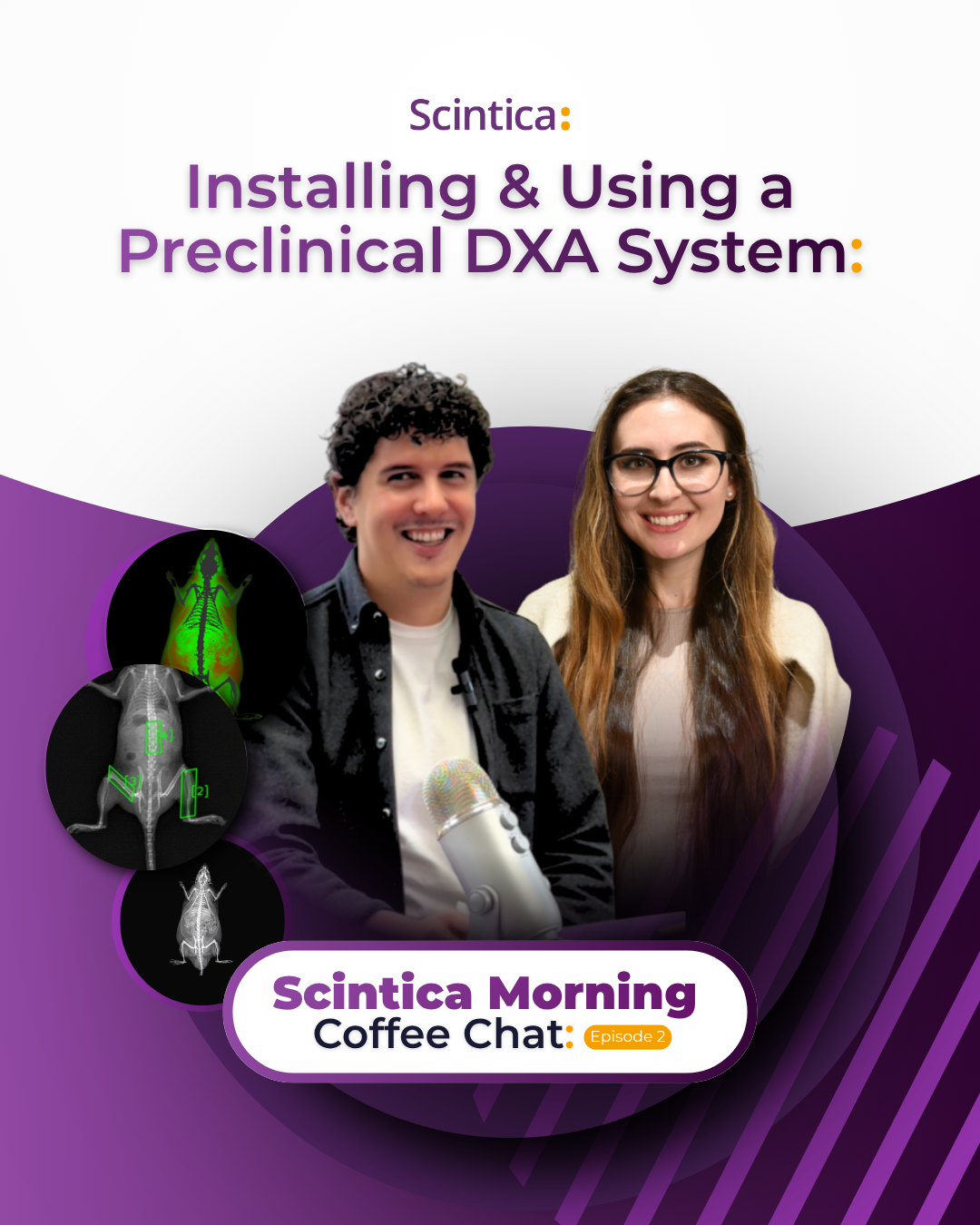
Scintica Morning Coffee Chat Episode 2: FAQ – Installing & Operating the iNSiGHT DXA System
https://youtu.be/Yi0lydfNs5I In this episode of Scintica Morning Coffee Chat, we examine the technical and operational requirements
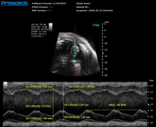
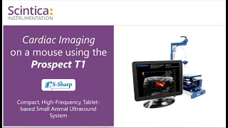
Clinical vs. Preclinical Ultrasound: Why Frequency Matters
Clinical vs. Preclinical Ultrasound: Why Frequency Matters View Paper Here Our Ultrasound SystemProspect
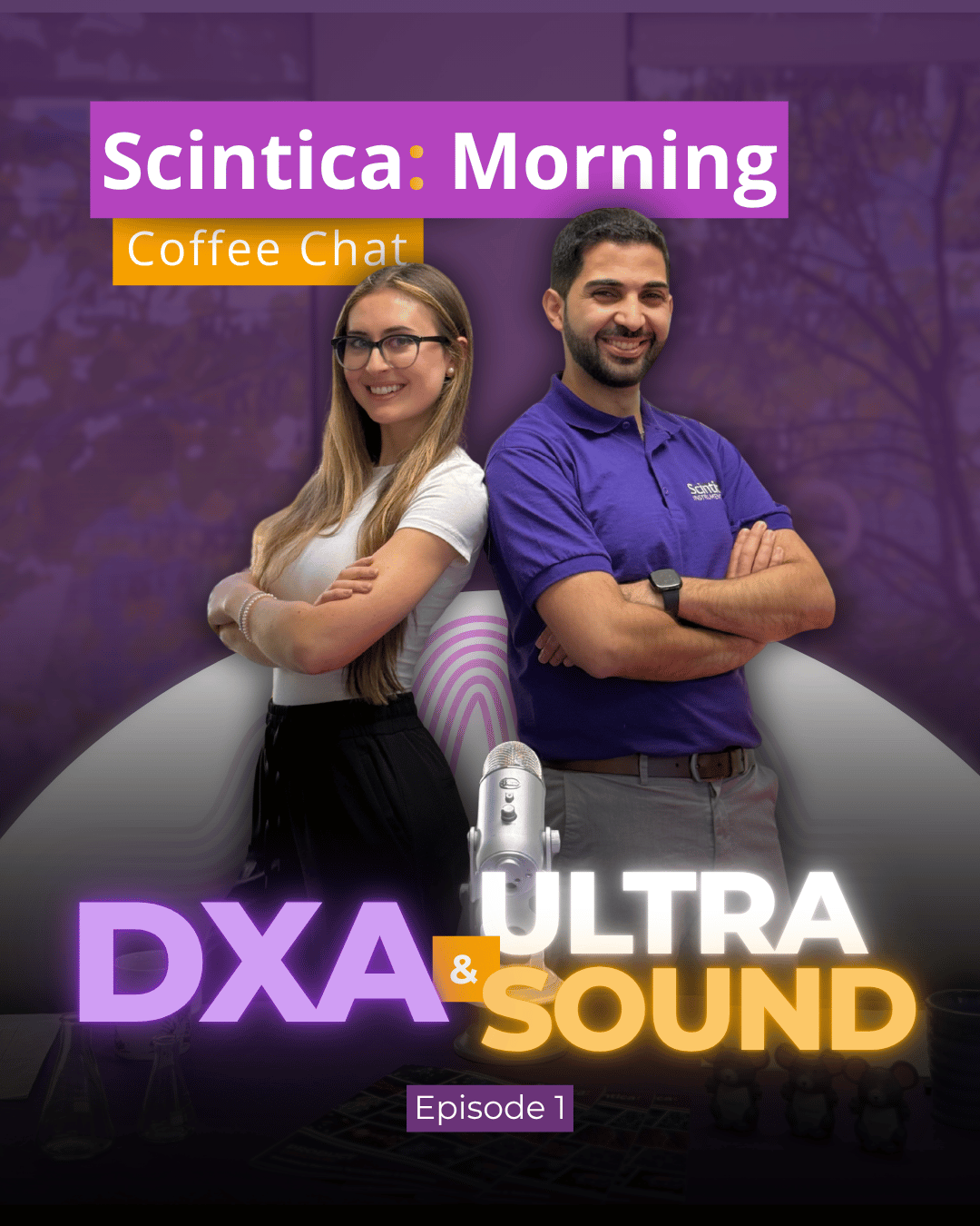
Scintica Morning Coffee Chat Episode 1: DXA & Ultrasound
https://youtu.be/KfbEW_b5Gpg?si=2xXwoxw-DuGtjq77 Join us on our first episode of the Scintica Morning Coffee Chat. This