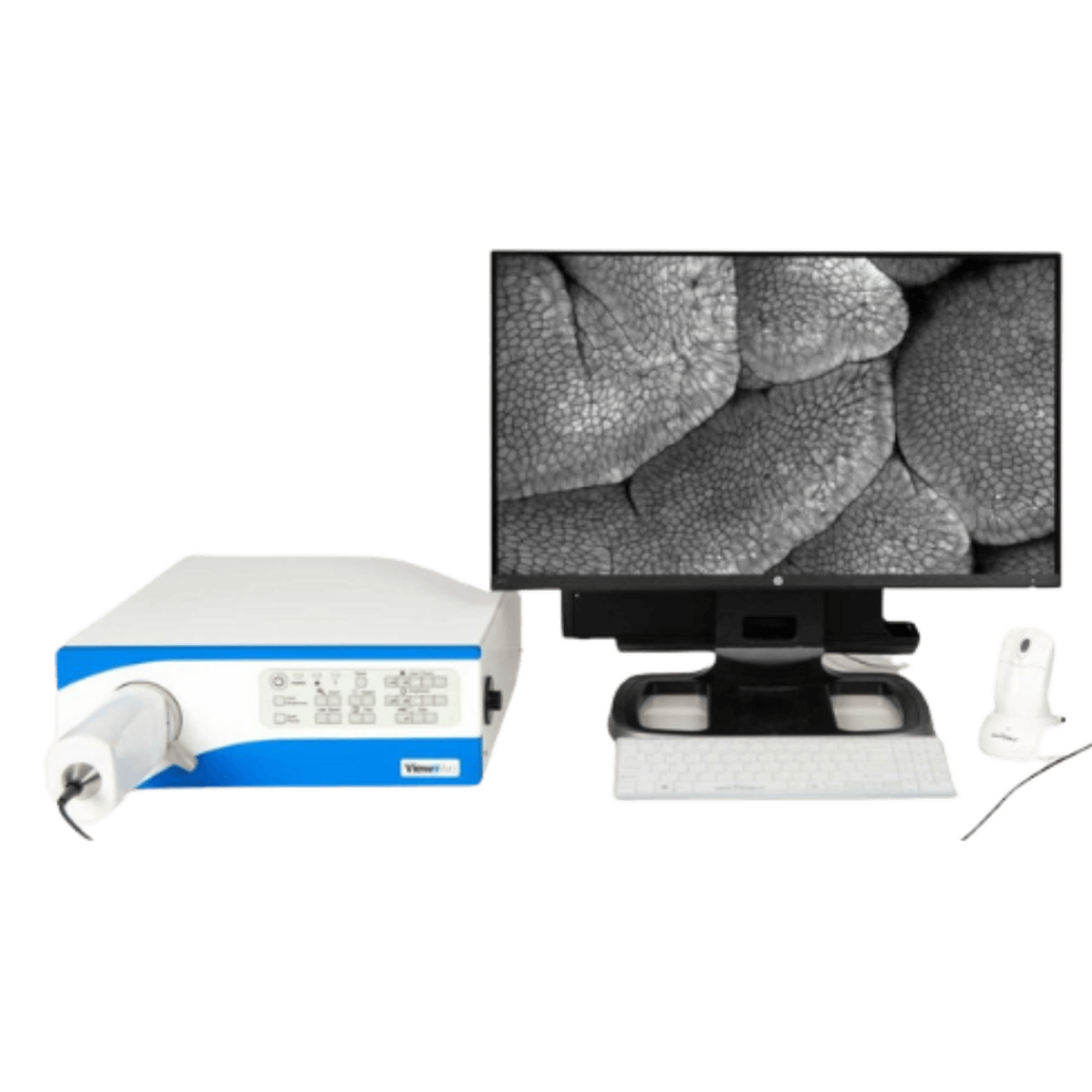

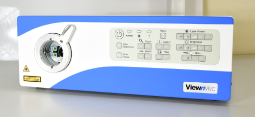
Confocal Processor
Optiscan Imager
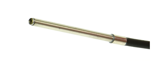
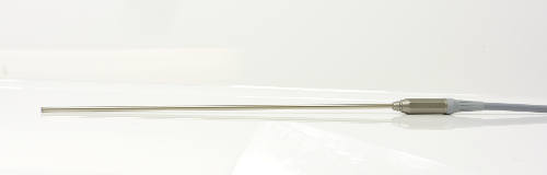
Probes
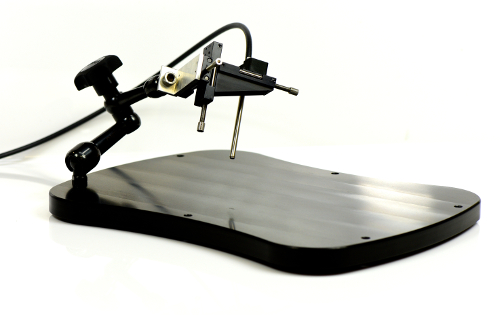
Animal Stage
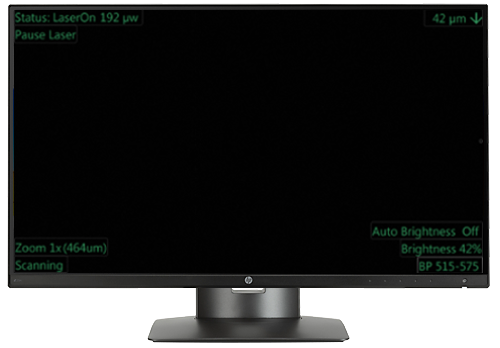
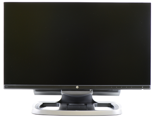
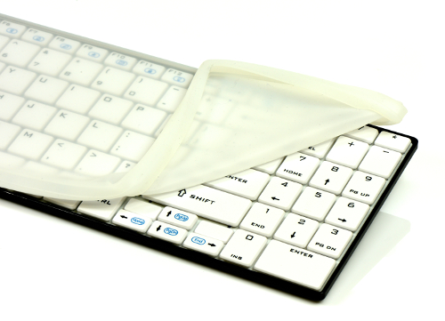
Computer Components
Footswitch
3D Software
Confocal Processor
Optiscan Imager
Probes

Animal Stage
Computer Components

Footswitch
3D Software
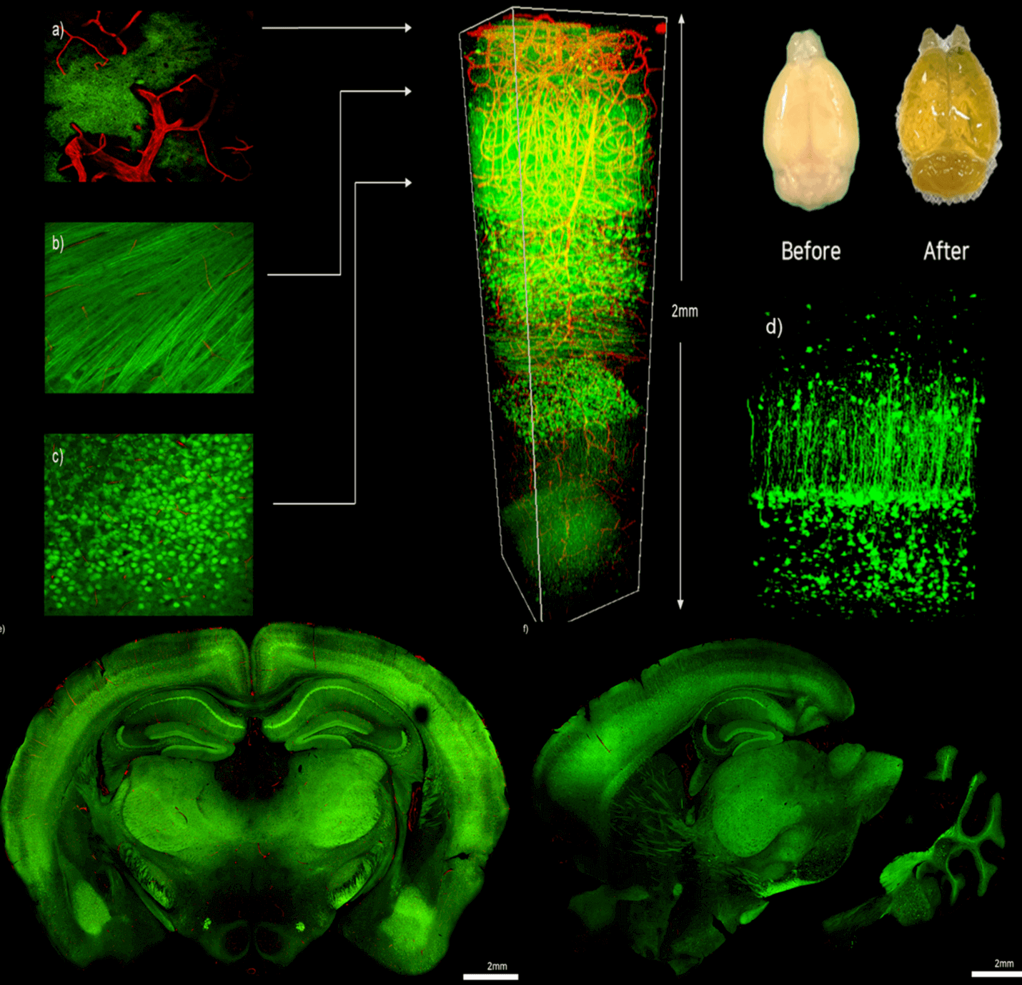
Leverage your research with IVIM Technology’s advanced imaging techniques for deep brain imaging
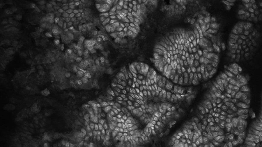
System Used In This Work: Intravital Microscopy (IVM) View Paper Here Introduction Our
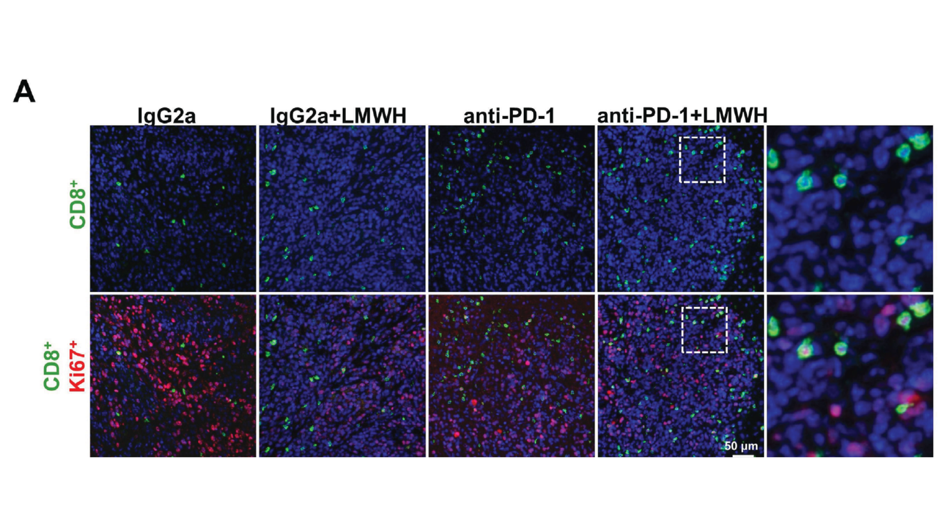
Low molecular weight heparin synergistically enhances the efficacy of adoptive and anti-PD- 1- based immunotherapy by increasing lymphocyte infiltration in colorectal cancer
Yibo Quan ,1 Jie He ,1,2 Qi Zou,1,2 Liuxi Zhang,1,2 Qihui Sun, 2
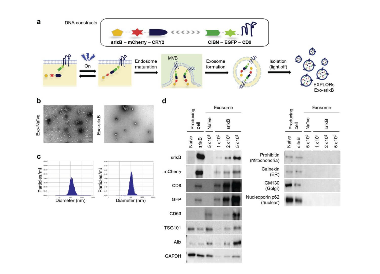
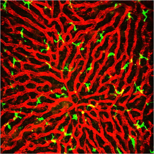
Exosome-based delivery of super-repressor IkBa ameliorates kidney ischemia-reperfusion injury
Exosome-based delivery of super-repressor IκBα ameliorates kidney ischemia-reperfusion injury Seonghun Kim1,7, Sul A
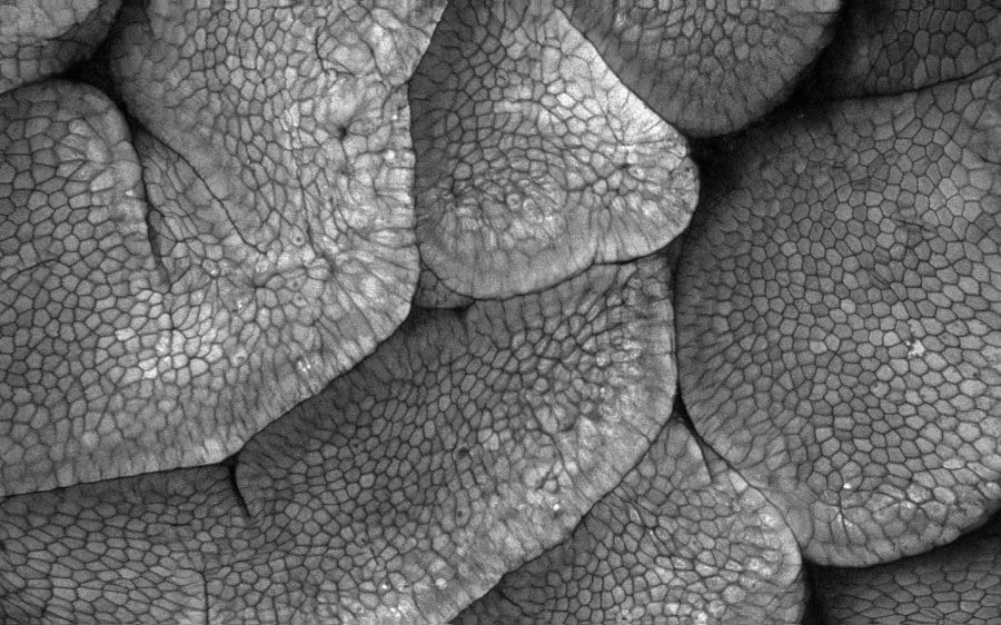
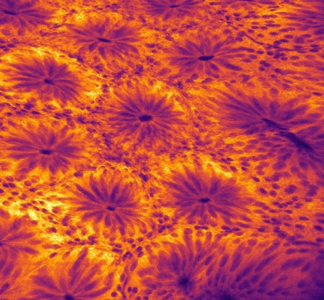
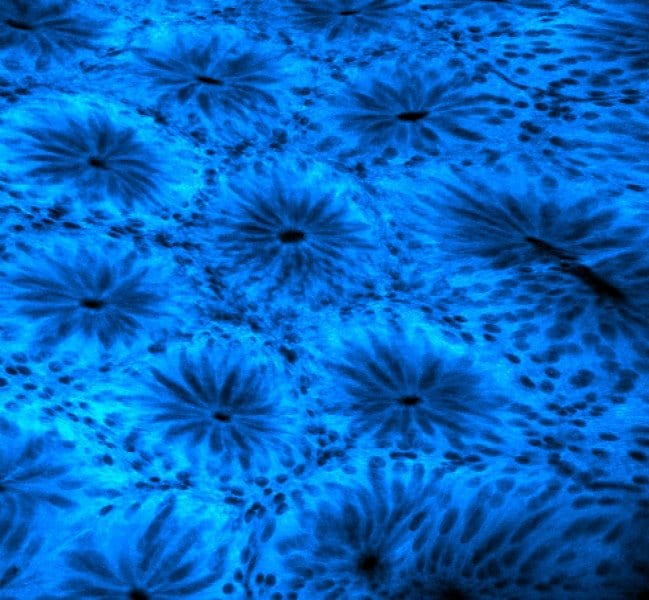
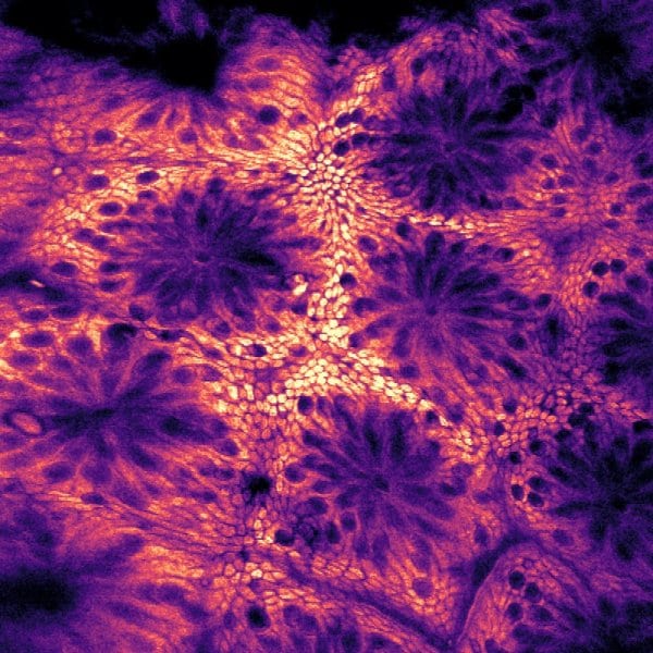
Application Note : Liver
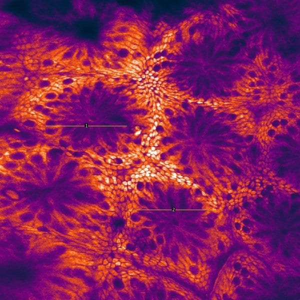
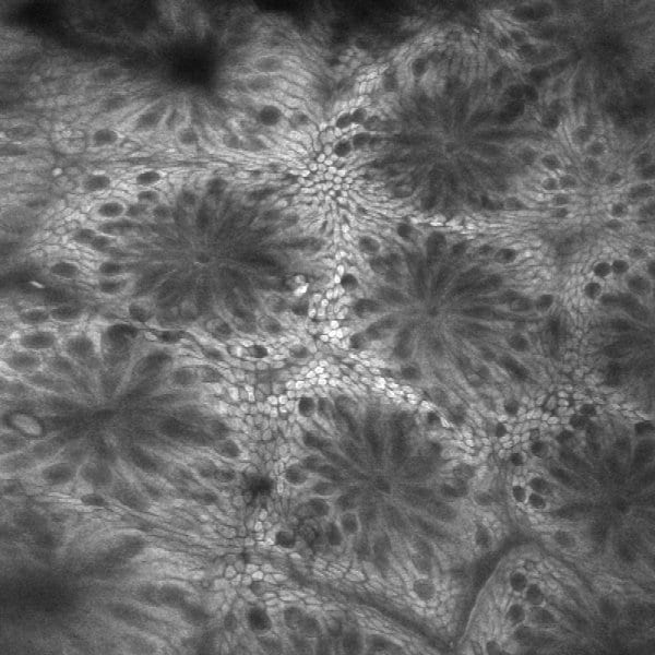
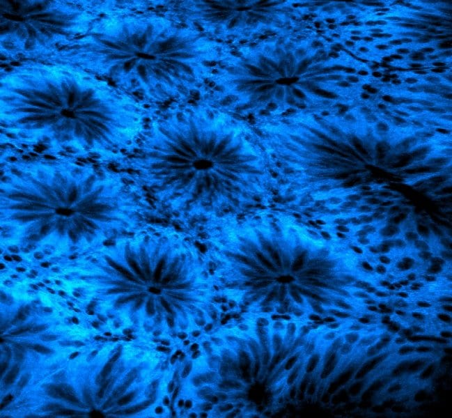
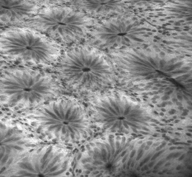
Intravital longitudinal imaging of hepatic lipid droplet accumulation in a murine model for