Preclinical Imaging:
Modalities Overview
Preclinical Imaging Modalities Used in Research
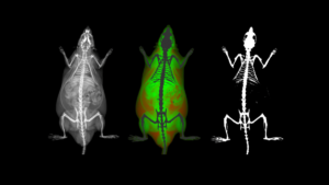
As we begin to explore the idea of multimodal imaging, let’s first start to explore the variety of preclinical imaging modalities that are most commonly used by researchers around the world. Many of the imaging modalities used in the clinic have been adapted for preclinical use with a focus on working with mice, rats, as well as some other species such as guinea pigs, hamsters, rabbits and even zebrafish, birds, ferrets, etc. These imaging modalities include MRI (magnetic resonance imaging), PET (positron emission tomography), CT (computed tomography), ultrasound (US), fluorescence (FLI), and DEXA (dual energy x-ray absorptiometry). While other imaging modalities are focused mostly on preclinical imaging to help understand the underlying mechanisms of disease, but are not necessarily directly translatable to the clinic, including bioluminescence (BLI) and intravital microscopy (IVM). While some still are newer to both clinical and preclinical research, but continue to advance over time, such as photoacoustic imaging (PAI).
Modality: Sensitivity vs. Resolution
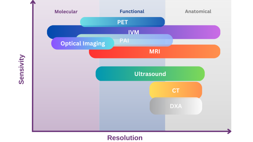
Each modality has its own strengths and weaknesses, and this may also include areas where they are more commonly used in preclinical research. However, in general, the use of more than one complimentary imaging modality would allow researchers to further elucidate molecular and biological mechanisms of disease, and to explore the response to a variety of therapeutic interventions. When considering an imaging modality, one must consider what is being imaged – i.e., soft tissue, bone, endogenous or exogenous contrast agents, etc. Additionally, resolution, radiation exposure, imaging time, and need for genetic manipulation or injection of additional agents are all factors that should be considered when designing an imaging experiment.
Finally, complimentary modalities should be selected as to not provide redundancy, but to augment the information to include as many of the following as possible – anatomical, morphological, functional and/or molecular data. Overall, the goal should be to provide a composite of complimentary information about a specific disease model over time; providing as much spatial and temporal information about a specific process as possible by developing a mechanistic understanding of the model at the whole body, organ, and/or cellular level.
Modality Summary
| Modality | Basis for Imaging | Contrast Mechanism | Spatial Resolution |
|---|---|---|---|
|
(MRI) |
Nuclear spins (i.e., 1H) are aligned in a magnetic field and then excited. The resulting RF signal is detected over time. |
Intrinsic contrast within the image is created by varying environments within which the nuclei (i.e., 1H) exist. Providing exquisite anatomical soft tissue images.
Extrinsic contrast agents can be used, such as gadolinium, 2+Mn, or iron oxide. These agents may be designed to detect functional or molecular targets. |
~100µm |
| Advantages | Limitations | Applications |
|---|---|---|
|
|
|
| Modality | Basis for Imaging | Contrast Mechanism | Spatial Resolution |
|---|---|---|---|
|
(CT) |
X-rays are passed through the sample. The resulting image is due to varying attenuation through the tissues. |
Intrinsic contrast within the image is created by varying attenuation of the x-rays. Providing very good anatomical images of hard structures such as bone, the lungs, with less definition in soft tissues.
Extrinsic contrast agents such as iodine may be used to detect functional targets. |
≤50µm |
| Advantages | Limitations | Applications |
|---|---|---|
|
|
|
| Modality | Basis for Imaging | Contrast Mechanism | Spatial Resolution |
|---|---|---|---|
|
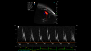
Ultrasound |
Sound waves are transmitted into the tissue, reflection of these waves is then detected. The acoustic impedance within tissue, and changes in the received signal are used to create the resulting images and spectrums. |
Intrinsic contrast within the image is created by varying acoustic impedance between various tissues providing very good anatomical soft tissue images.
Extrinsic contrast agents can be used, such as microbubbles or nanoparticles. These agents may be designed to detect functional or molecular targets. |
~30-50µm |
| Advantages | Limitations | Applications |
|---|---|---|
|
|
|
| Modality | Basis for Imaging | Contrast Mechanism | Spatial Resolution |
|---|---|---|---|
|
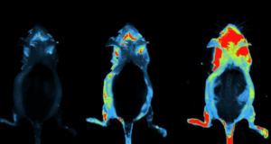
(OI) |
Light emission is detected by very sensitive detectors. The source of the light varies between bioluminescence and fluorescence. |
Bioluminescence imaging requires the expression of a specific enzyme within the cells of interest, with the substrate being injected at the time of imaging. The resulting enzymatic reaction results in the release of photons.
Fluorescence imaging requires either intrinsic fluorophores or injected imaging agents. These fluorophores must be excited using a defined wavelength, while the emitted photons are detected to generate the image.
Both of these may be used to detect functional or molecular signals. |
~1mm |
| Advantages | Limitations | Applications |
|---|---|---|
|
|
|
| Modality | Basis for Imaging | Contrast Mechanism | Spatial Resolution |
|---|---|---|---|
|
(PAI) |
Specific wavelengths of light are used to excite specific chormophores which then create an acoustic effect which is detected to generate an image. |
Intrinsic molecules, such as hemaglobin, may result in a photoacoustic signal. While other imaging agents may be injected to acquire signals from a specific target.
Depending on the agents used functional and molecular signals are most common, however some anatomical information can be obtained. |
~300um |
| Advantages | Limitations | Applications |
|---|---|---|
|
|
|
| Modality | Basis for Imaging | Contrast Mechanism | Spatial Resolution |
|---|---|---|---|
|
(IVIM) |
Defined wavelengths of light are used to excite specific molecules. Images are acquired using microscopic objectives. |
Some native intrinsic molecules provide a signal, such as collagen, or cells which have been genetically modified to express a fluorophore are incorporated into a model. In other situations, molecules or cells tagged with a specific fluorophore are injected at the time of imaging.
Confocal or Two-Photon techniques are used to excite the specific fluorophores, while microscope objectives are used to acquire the images within a specific field of view. |
~1µm |
| Advantages | Limitations | Applications |
|---|---|---|
|
|
|
| Modality | Basis for Imaging | Contrast Mechanism | Spatial Resolution |
|---|---|---|---|
|
(DEXA) |
X-rays of two specific energy levels are passed through the sample. The resulting image is due to varying attenuation through the tissues.
Specific equations are used to determine if each pixel belongs to the category of bone or soft tissue. Further, the soft tissue pixels are determined to be fat or lean mass. Additional information about the properties of the bone is also provided. |
Intrinsic contrast within the image is created by varying attenuation of the x-rays. Providing very good anatomical images of hard structures such as bone, the lungs, with less definition in soft tissues. |
~100µm for DEXA image
~30µm for highest magnification digital radiography image |
| Advantages | Limitations | Applications |
|---|---|---|
|
|
|
Modality Comparision
| Modality | Resolution | Advantages | Limitations | Applications |
|---|---|---|---|---|
|
MRI |
~100µm |
– Non-ionizing radiation – Whole body imaging is possible – Excellent soft tissue contrast – Clinical translation |
– Some systems are expensive and high maintenance costs **Note M-Series compact MRI does not have these limitations – No bone contrast |
– Cancer Biology – Neurology – Organ Imaging – Cardiac Imaging – Contrast Agent Imaging |
|
CT |
≤50µm |
– Whole body imaging is possible – Excellent bone imaging – Clinical translation |
– Ionizing radiation – Low soft tissue contrast |
– Bone Imaging – Anatomical Reference for Other Imaging Modalities |
|
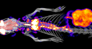
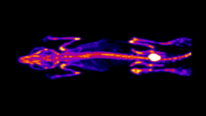
PET |
~1mm |
– Whole body imaging is possible – High sensitivity to agents used – Quantifiable data – Wide variety of applications, based on imaging agents used – Clinical translation |
– Radioactive agents are required – Specialized infrastructure required – Proximity to cyclotron, more important for short half-life molecules |
– Cancer Biology – Neurology – Cardiac Imaging – Pharmacokinetics and Pharmacodynamic Imaging |
|
Ultrasound |
~30-50µm |
– High temporal and spatial resolution – Good soft tissue contrast – Non-ionizing radiation – Clinical translation |
– Depth of penetration vs resolution – Operator dependent |
– Cancer Biology – Organ Imaging – Cardiac Imaging – Vascular Imaging – Developmental Biology – Contrast Agent Imaging |
|
Optical |
~1mm |
– Non-Ionizing radiation – Whole body imaging is possible – High throughput – Wide variety of applications depending on agent used – Ease of use |
– Limited tissue penetration for some photons – Low spatial resolution – Semi-quantitative |
– Cancer Biology – Cell Trafficking – Gene Expression |
|
Photoacoustic |
~300um |
– Whole body imaging is possible – Penetration depth – Endogenous and exogenous contrast |
– Anatomical & structural information |
– Cancer Biology – Neurology – Developmental Biology – Contrast Agent Imaging – Angiograph |
|
Intravital Microscopy |
~1µm |
– Multiplex imaging capabilities – Microscopic resolution – Dynamic imaging allowing for live cell and molecule tracking – Wide variety of applications |
– Depth of penetration – Small field of view – Surgical preparation to access imaging target |
– Cancer Biology – Neurology – Cardiac Imaging – Vascular Imaging – Organ Imaging – Musculoskeletal Imaging – Cell Tracking |
|
DEXA/DXA |
~100µm for DEXA ~30µm for highest magnification digital radiography image |
– Rapid scan times – Whole body imaging is possible – Bone mineral density/content measurements provided – Lean vs. fat mass measured – Very low ionizing radiation level per scan, allowing for longitudinal imaging – Clinical translation |
– Images are 2D – Low soft tissue contrast |
– Bone Imaging – Body Composition Measurements – Digital Radiography |
Next Modules
Preclinical Imaging Modalities
Available Now
As we begin to explore the idea of multimodal imaging, let’s first start to explore the variety of preclinical imaging modalities that are most commonly used by researchers around the world.
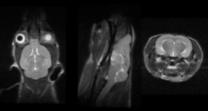
Modality: MRI
Available Now
MRI is considered the gold standard in soft tissue imaging, both in the clinic on patients and by researchers on a wide variety of preclinical imaging subjects.
Modality: CT
Available Now
Computed Tomography (CT) is one of the most commonly used clinical imaging techniques, next to perhaps ultrasound.
Modality: PET
Available Now
Nuclear imaging modalities include positron emission tomography (PET) as well as single photon emission computed tomography (SPECT), which both detect gamma radiation emitted from a radionucleotide injected into an imaging subject.
Modality: Ultrasound
Available Now
Ultrasound (US) imaging is one of the most commonly used diagnostic techniques clinically, and is widely used in preclinical imaging as well. It is a safe, non-invasive, and relatively inexpensive technology compared to some of the other available imaging modalities.
Modality: Optical
Available Now
When discussing Optical Imaging techniques in this series, the meaning is to cover bioluminescence (BLI) and fluorescence (FLI) imaging. T
Modality: Photoacoustic
Available Soon
Photoacoustic (PAI) imaging is a technology which combines the sensitivity of optical imaging with the depth of penetration and resolution of ultrasound imaging.
Modality: Microscopy
Available Soon