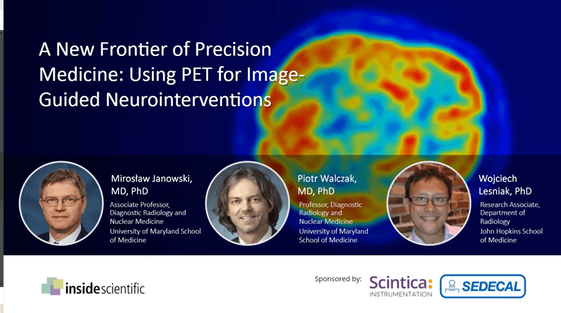
WEBINAR: A New Frontier of Precision Medicine: Using PET for Image-Guided Neurointerventions
Thursday, May 7 2020
In this webinar sponsored by Scintica Instrumentation, Dr. Piotr Walczak, Dr. Mirosław Janowski and Dr. Wojciech Lesniak addressed this challenge and discussed why advanced imaging is essential to perform image-guided neurointerventions.
Mice are by far the most frequently used animal for modeling disease and developing therapeutic strategies including neurointerventions. However, due to its anatomical and physiological barriers, the brain is a difficult target for delivery of therapeutic agents. Systemic administration is plagued with marginal brain accumulation and high risk of off-target side effects.
In this webinar sponsored by Scintica Instrumentation, Dr. Piotr Walczak, Dr. Mirosław Janowski and Dr. Wojciech Lesniak addressed this challenge and discussed why advanced imaging is essential to perform image-guided neurointerventions.
First, Dr. Janowski provided rationale as to how imaging can be used to better understand how therapeutic agents are delivered to the brain and subsequently cleared. Next, Dr. Walczak reviewed methodological and technological advances for improving precision and reproducibility of brain targeting in mice based on MRI and two-photon microscopy. Finally, Dr. Lesniak presented recently-published results using ARGUS PET/CT to quantify intra-artrial delivery of antibodies, nanobodies and poly(amidoamine) dendrimers.
Key discussion topics included:
- Why advanced imaging is essential to perform image-guided neurointerventions
- Why we need to visualize not only penetration of therapeutic agents to the brain but also their clearance
- How image-guided procedures can be used to visualize and optimize delivery of therapeutic agents to the brain
About the Speaker (s)
Mirosław Janowski, MD, PhD
Associate Professor, Diagnostic Radiology and Nuclear Medicine
School of Medecine
University of Maryland
Dr. Janowski’s background in clinical neurosurgery, neuroscience and imaging provides an excellent basis for comprehensive tackling of central nervous system diseases. Early in his career he recognized a peculiarity of the brain and spinal cord, which are hidden behind the skull and blood brain barrier. We are witnessing much progress in the treatment of many diseases, while the therapeutic outcomes of brain disorders lag behind. Therefore, his current focus is on the development of methods for effective delivery of therapeutic agents to the brain, and advanced imaging is pivotal to proper understanding of therapeutic agent kinetics including delivery and clearance from the brain.
Piotr Walczak, MD, PhD
Professor, Diagnostic Radiology and Nuclear Medicine
School of Medicine
University of Maryland
Dr. Walczak received his medical degree from the Medical University of Warsaw and PhD in regenerative medicine from the University of Warmia and Mazury in Poland followed by postdoctoral training at the University of South Florida and the Johns Hopkins University. He served on a faculty at Hopkins until 2019 when he joined UMB Department of Diagnostic Radiology and Nuclear Medicine. His research is focusing on developing advanced tools for guiding brain repair strategies. His lab utilizes animal models and multimodal imaging at various scales, including MRI, PET and intravital microscopy, to interrogate mechanisms and improve efficacy of therapeutic agents, such as stem cells, macromolecules and gene therapeutics.

Wojciech Lesniak, PhD
Research Associate
Department of Radiology
Johns Hopkins School of Medicine
Dr. Wojciech Lesniak earned a M.Sc. in Chemistry (1997) and a Ph.D. in Bioinorganic Biomedical Chemistry (2001) at the University of Wroclaw (Wroclaw, Poland). Afterwards, Dr. Lesniak completed post-doctoral training at the University of Michigan (2004) in the Department of Chemistry and the Center of Nanotechnology for Medicine. He then worked as an Oncology Instructor at the Roswell Park Cancer Institute (2005-2010) and a Research Associate at the Wayne State University (2010-2011). Since 2012, he has been working at the Johns Hopkins School of Medicine as a Research Associate in the Department of Radiology and, in 2019 was promoted to serve as a Director of the Radiometabolite Laboratory at PET center. He has been involved in multidisciplinary research related to development and evaluation of low molecular weight scaffolds, nanoparticles and theranostic agents for targeted cancer imaging and therapy. He authored 55 peer-reviewed scientific papers, book chapters and a few patents.