(May 17, 2021) Introducing the Newton 7.0 Optical Imaging System: The Modality, System Features and Applications
Overview:
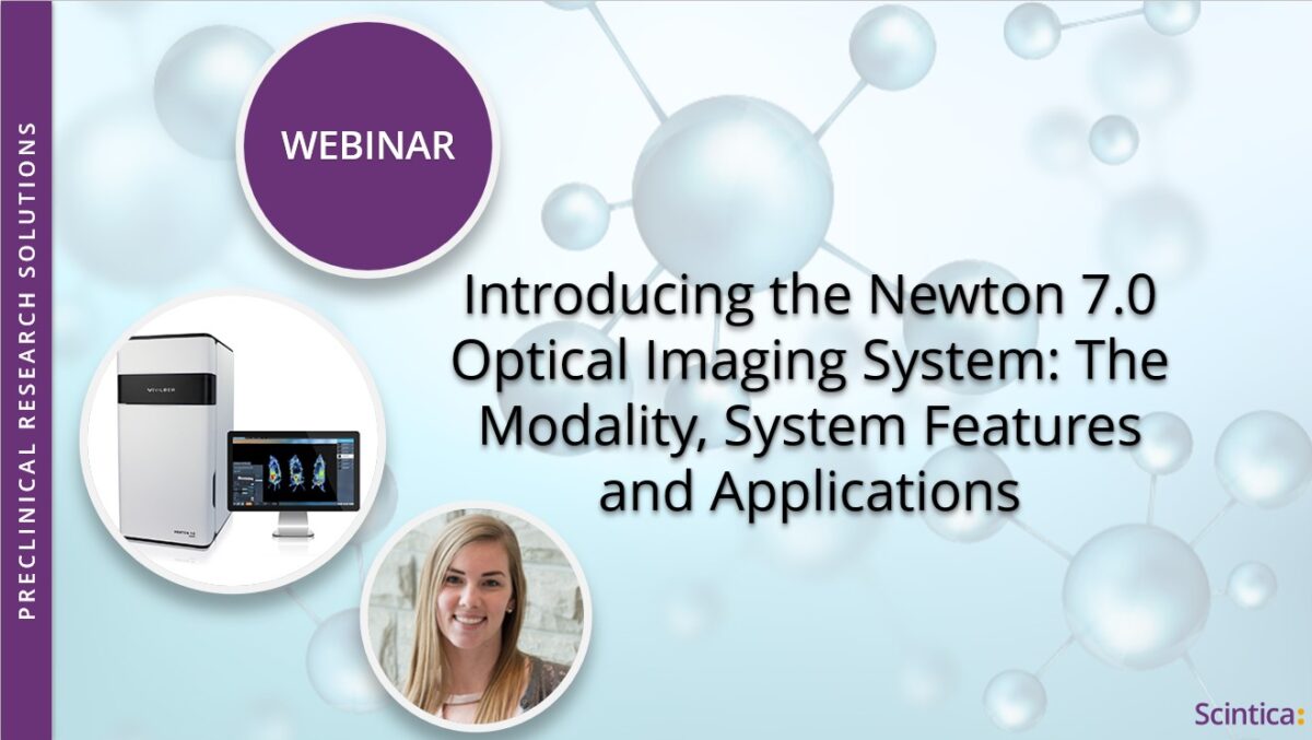
In this webinar, Katie reviewed how optical imaging works and provided some common examples of how it’s used within preclinical research. She introduced the Newton 7.0 FT500, an optical imaging system manufactured by Vilber that included bioluminescence, fluorescence, and 3D tomography capabilities. She concluded the webinar by going through some of the most common questions/considerations that come up for those looking to add optical imaging capabilities to their lab.
The Newton 7.0 is a highly sensitive optical imaging system dedicated to preclinical imaging of small animals in vivo, and may also be used on a variety of in vitro and ex vivo samples. It combines the best optics and animal handling features for high-quality image data and quantification. The Newton 7.0 system is capable of bioluminescence, fluorescence as well as 3D tomographic imaging.
The system is:
- User-friendly
- Does not require any radiation to acquire images
- Is non-invasive, allowing for longitudinal studies
- Allows for up to 5 mice or 3 rats to be imaged simultaneously
The Newton 7.0 is highly sensitive and can be used in a wide variety of research applications. Various bioluminescent reporters like firefly luciferase and many fluorescent molecular reagents can be used to visualize and track tumors, monitor disease or inflammation development, target molecules to nanoparticles or follow biodistribution and pharmacokinetics noninvasively.
Whether you are just exploring the idea of adding optical capabilities to your lab or you’ve been imaging for years, you won’t want to miss the opportunity to learn about this cool new technology.
Learning objectives:
- How does optical imaging work?
- Common applications of preclinical optical imaging
- A product overview: Newton 7.0 FT500
- What makes the system unique?
- How to add optical imaging capabilities to your lab?
In collaboration with The Keenan Research Center for Biomedical Science, Toronto
About the Speaker (s)

Katie Parkins, Ph.D.
Preclinical Imaging Specialist, Scintica
Katie Parkins holds a Ph.D. from the University of Western Ontario in Medical Biophysics. Throughout her research training, she was focused on the development and application of novel molecular imaging tools to monitor and treat disease, specifically, cancer. Additionally, Katie completed a Postdoctoral Fellowship at Johns Hopkins University where she used multimodal imaging to investigate the molecular mechanisms that may determine immunotherapy response. In her present role as Preclinical Imaging Specialist at Scintica Instrumentation, Katie supports our customers in understanding the products offered and how these instruments help to meet their research needs.