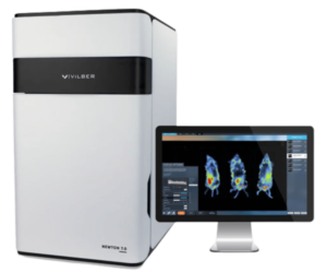
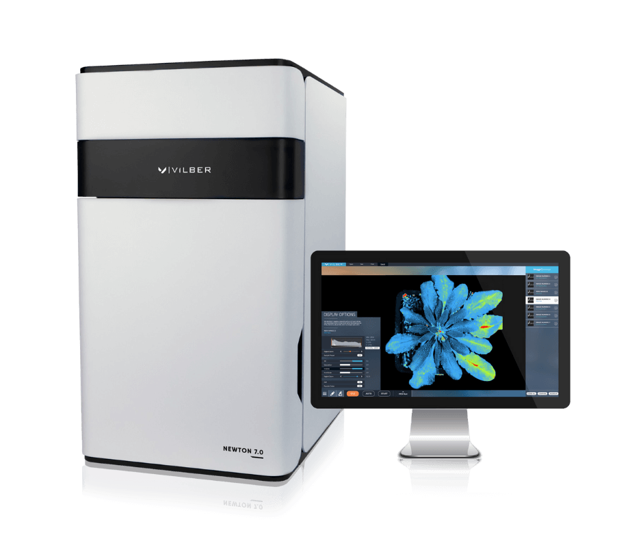
Newton 7.0
In Vivo Optical, Fluorescence & Bioluminescence, Imaging


System Overview
The Newton 7.0 is a cutting-edge optical imaging system that offers the versatility to perform bioluminescence, fluorescence, and 3D tomographic imaging in a single device. Its user-friendly interface and advanced features make it ideal for in vivo, ex vivo, and in vitro imaging applications, as well as simultaneous imaging of multiple specimens.
The system features a state-of-the-art 4.6 megapixel CCD camera that boasts one of the largest apertures in the market. This camera provides excellent sensitivity for a variety of luciferase enzymes and fluorophores commonly used in preclinical research, allowing for fast and efficient signal acquisition. The intuitive workflow and user-friendly software are optimized for multi-user use, saving valuable time in longitudinal studies.
What Makes the NEWTON Optical Systems Stand Out?
Features & Benefits
The Newton 7.0 is an innovative optical bioluminescence, fluorescence, and 3D tomographic imaging system designed with the user in mind.
State-of-the-art Camera Technology
- Scientific grade 0 16-bit CCD
- 4.6 MP Native Resolution
- -90oC Absolute Cooling
- f/0.7 Lens Aperture
- 10 MP Image Resolution
- 4.8 Optical Density
Powerful Fluorescent Excitation
The Newton 7.0 features eight excitation channels across the visible and near-infrared spectrums. Two powerful Laser Class II arrays tightly control the illumination light, delivering a direct, high-intensity light.
Motorized Darkroom with Adjustable Field-of-View
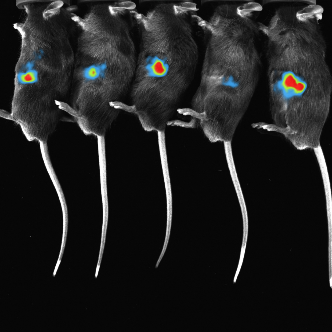
Vilber’s intelligent darkroom architecture allows for a fully motorized movement of the camera (Z-axis) and animal pad (X/Y axis) to move through both the macro imaging FOV (6x6cm) to the full FOV (20x20cm) for imaging up to 5 mice.
Full Spectrum Tunability
Powerful with 8 excitation channels and 8 emission filters that cover the complete spectrum from blue to infrared.
Hard-coated narrow bandpass filters are used for the collection of emission wavelengths to reduce cross talk between signals, allowing for seamless imaging of all of the most commonly used fluorophores.
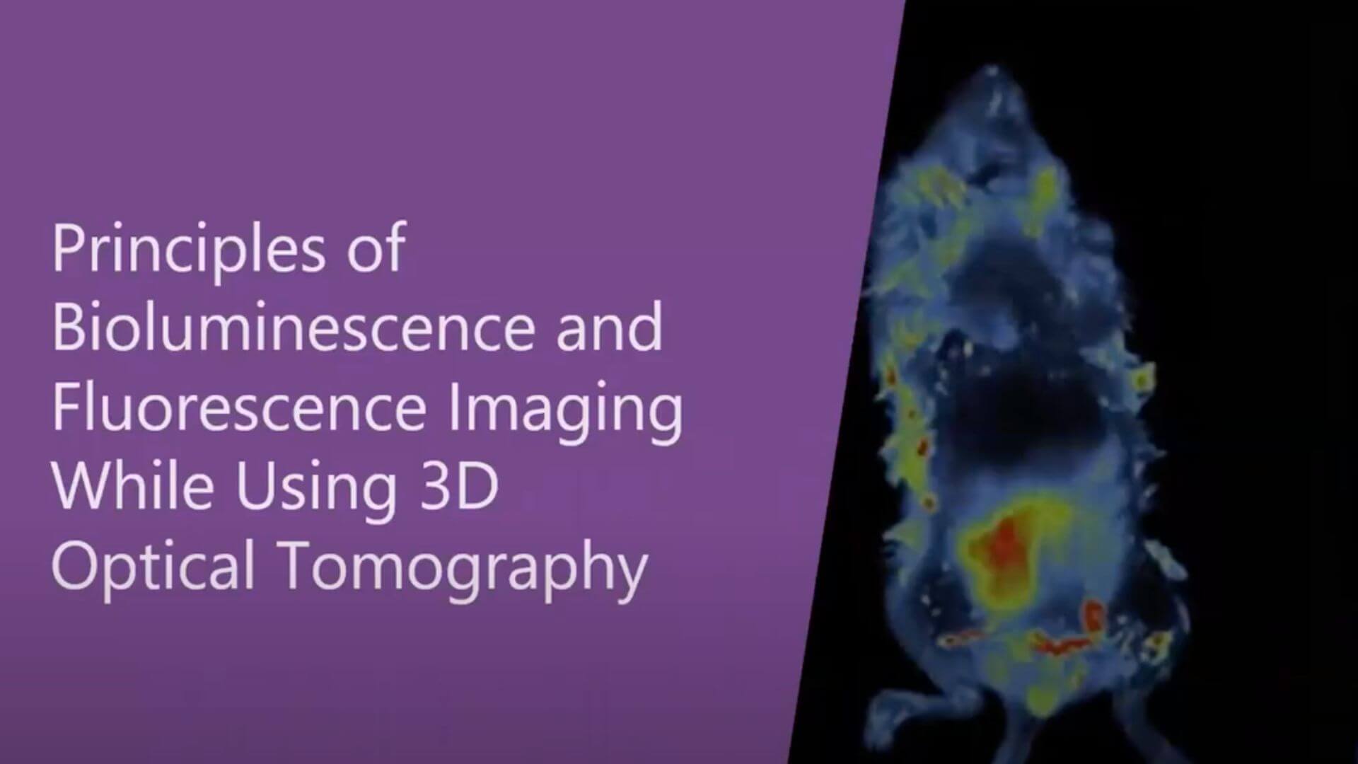
3D Optical Tomography
An integrated 3D tomography module allows for bioluminescent signals to be reconstructed in 3D and overlaid within a topographical model of the imaging subject.
The digital organ and bone library allows for the superimposition of the mouse organs and bones onto the topographical model to better understand anatomical and deeper tissue structures.
License-Free Acquisition and Analysis Software
The user-friendly software comes with unlimited licenses and free updates.
New and experienced users can quickly image their subjects using factory default acquisition protocols or create custom acquisition protocols.
Fully GLP and CFR21-compliant data can be exported in 16-bit .tiff or 8-bit .jpg formats.
In the analysis module, save ROI and analysis templates for high throughput analysis of up to 10 images side by side.
Imaging Modes
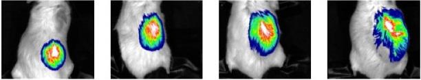
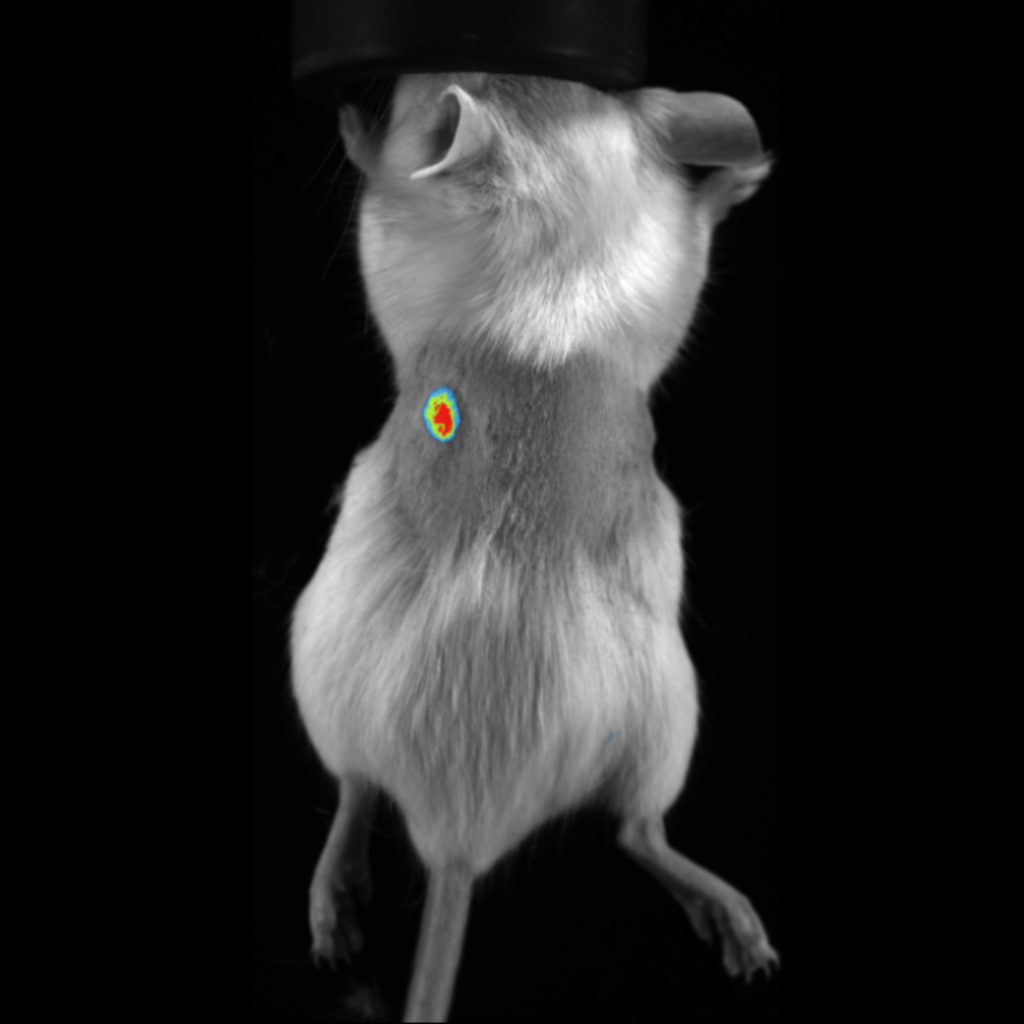
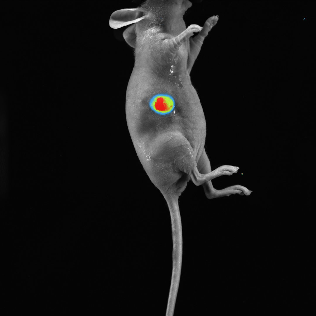
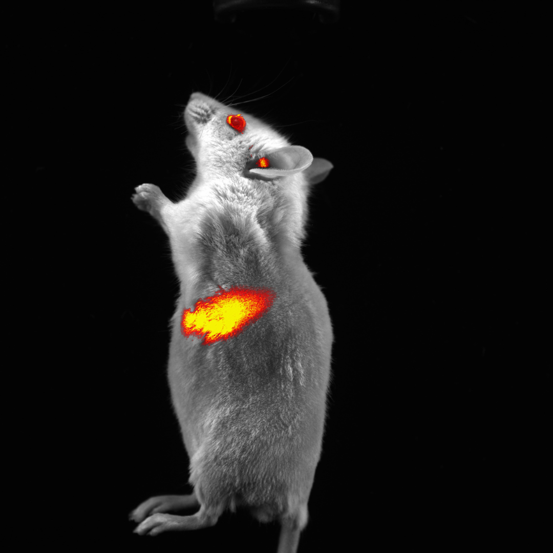
Fluorescence Imaging
Fluorescence imaging
Subcutaneous tumor expressing mCherry
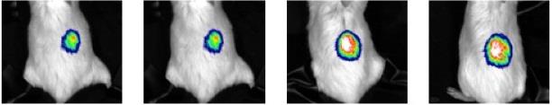
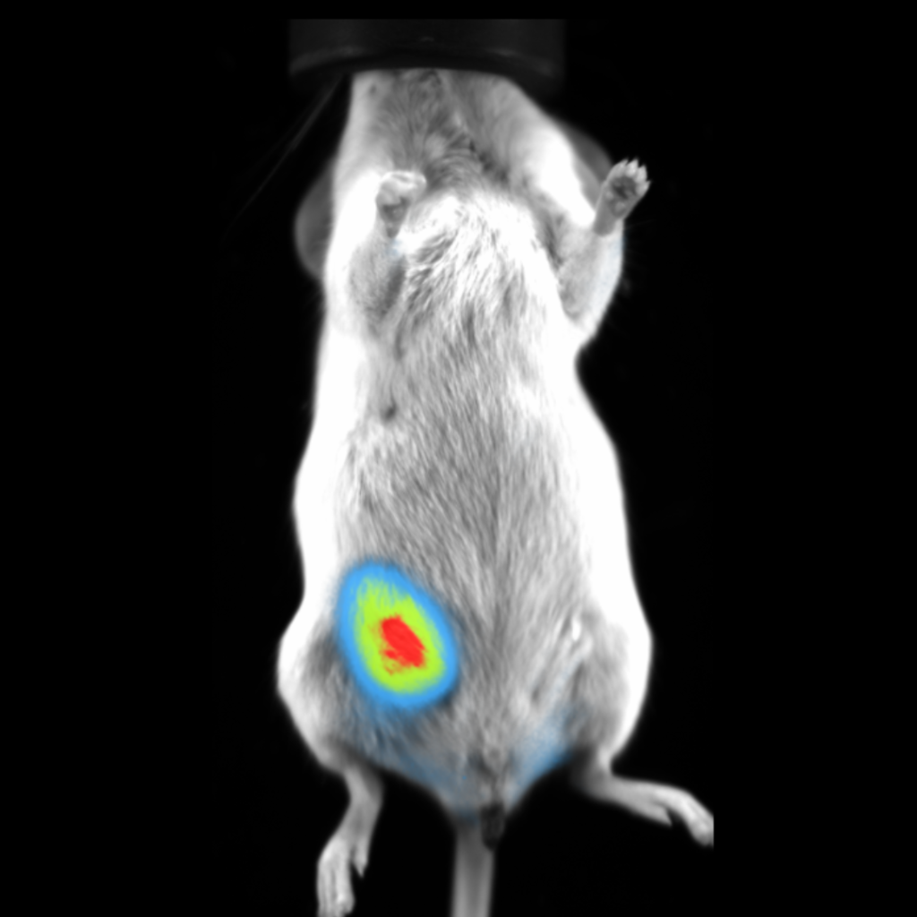
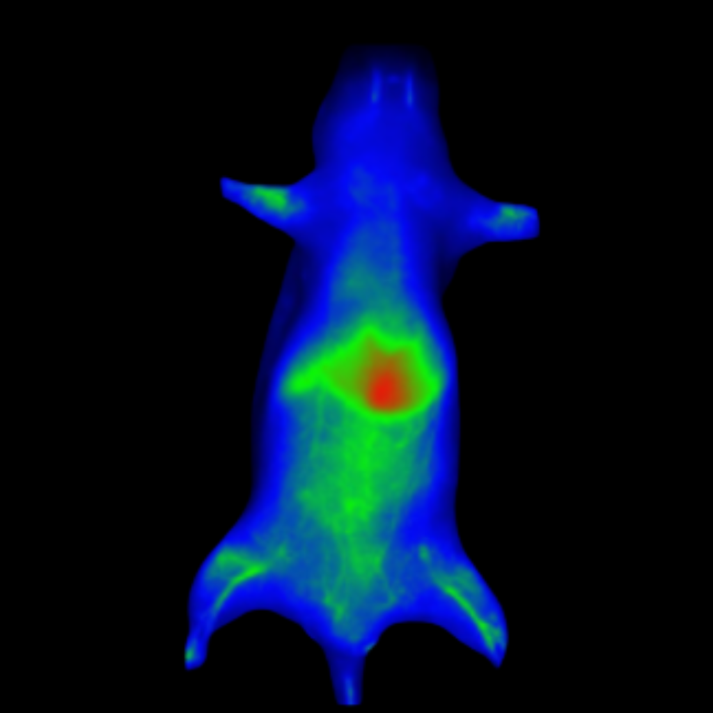
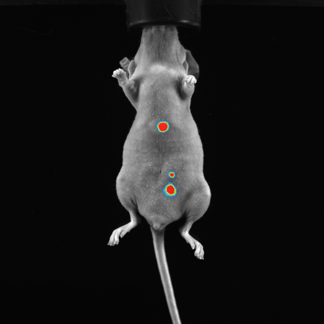
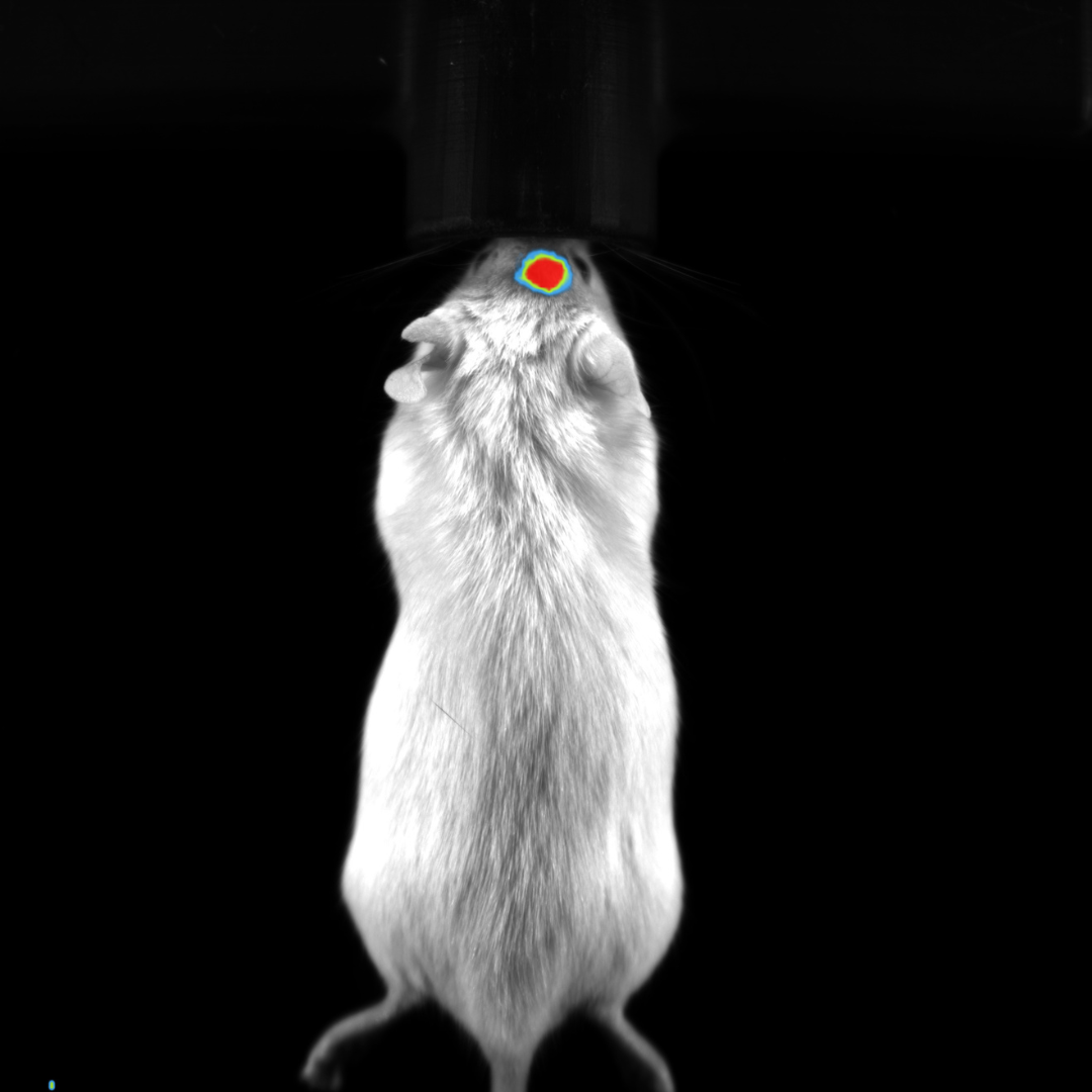
Bioluminescence Imaging
Bioluminescence imaging
Subcutaneous tumor expressing firefly luciferase
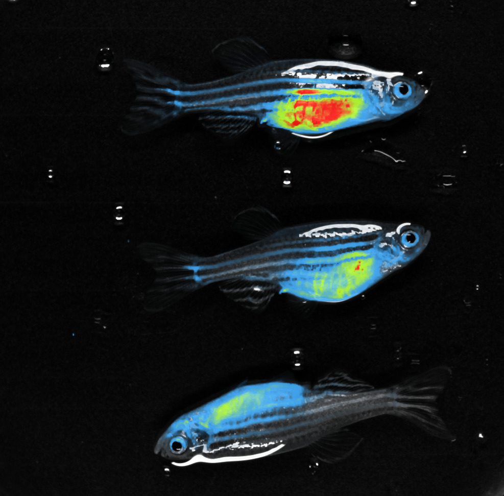
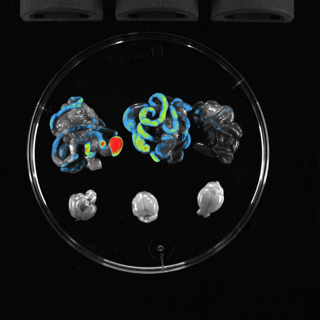
Multispectral In Vivo Imaging
Multispectral Imaging
Multispectral imaging
Longitudinal Imaging
- Images that are acquired at different time points can be arranged to form a longitudinal image sequence.
For example, a time series could be constructed from images acquired on different days following an experimental treatment. - Software then compares the image data throughout the experimental treatment.
Models & Specifications

NEWTON 7.0
MODELS BT100 & BT500-
In Vivo / In Vitro Optical imaging platform
-
Bioluminescence Detection
-
3D BLI Optical Tomography
-
Mice and Rats
NEWTON 7.0
MODELS FT100 & FT500-
In Vivo / In Vitro Optical imaging platform
-
Bioluminescence Detection
-
3D BLI Optical Tomography
-
VIS and NIR-1 Fluorescence
-
Mice and Rats
NEWTON 7.0
BIO-
In Vivo / In Vitro Optical imaging platform
-
Bioluminescence Detection Fentogram
-
Fluorescence Detection Picogram
- Camera
All Models
- 16-bit Scientific Grade CCD Camera
- Cooling: -90°C absolute
- Cooling: -120°C Delta
- Lens
All Models
- Proprietary V.070 – Fixed
- Focal Length Motorized lens
- Aperture: f/0.7
- Resolution
All Models
- Resolution: 2160×2160
- Monochrome & Color Imaging
- Emission
All Models
- 11-Position motorized filter wheel for all models
- 8 Narrow Band-pass filters included as standard 500 / 550 / 600 / 650/ 700/ 750 / 800 / 850 nm
Excitation
| BT100 | BT500 | FT100 | FT500 | BIO | |
|---|---|---|---|---|---|
|
White-Light |
Dual EPI-White Light LED Panels |
Dual EPI-White Light LED Panels |
Dual EPI-White Light LED Panels |
Dual EPI-White Light LED Panels |
Dual EPI-White Light LED Panels |
|
Fluorescence |
Upgradeable to Fluorescence |
Upgradeable to Fluorescence |
8 Fluorescent Channels Included 440 / 480 / 540 / 580 / 640 / 680 / 740 / 780 nm |
8 Fluorescent Channels Included 440 / 480 / 540 / 580 / 640 / 680 / 740 / 780 nm |
8 Fluorescent Channels Included 440 / 480 / 540 / 580 / 640 / 680 / 740 / 780 nm |
Emission
| BT100 | BT500 | FT100 | FT500 | BIO | |
|---|---|---|---|---|---|
|
Filter Wheel |
11-position Motorized Filter Wheel |
11-position Motorized |
11-position Motorized Filter Wheel |
11-position Motorized Filter Wheel |
11-position Motorized Filter Wheel |
|
Emission Filters |
4 Narrow Band-pass filters included for BLI Tomography: |
4 Narrow Band-pass filters included for BLI Tomography: 500/550/600/650nm |
8 Narrow Band-pass filters included: |
8 Narrow Band-pass filters included: |
8 Narrow Band-pass filters included: |
Darkroom
| BT100 | BT500 | FT100 | FT500 | BIO | |
|---|---|---|---|---|---|
|
Motorization |
|
|
|
|
|
|
Animal Handling |
|
|
|
|
|
- Animal Handling - All models
Heated Mouse Bed (+37°C) (included)
Animal breathers (included)
(Not Included with Newton 7.0 BIO)
Accessories / Add-ons
| BT100 | BT500 | FT100 | FT500 | BIO | |
|---|---|---|---|---|---|
|
Monitorization |
Fixed Camera Fixed Animal Stage |
Z-Axis Motorized Camera X/Y-Axis Motorized Animal Stage |
Fixed Camera Fixed Animal Stage |
Z-Axis Motorized Camera X/Y-Axis Motorized Animal Stage |
Z-axis Motorized Camera 15° Tilting Sample Stage |
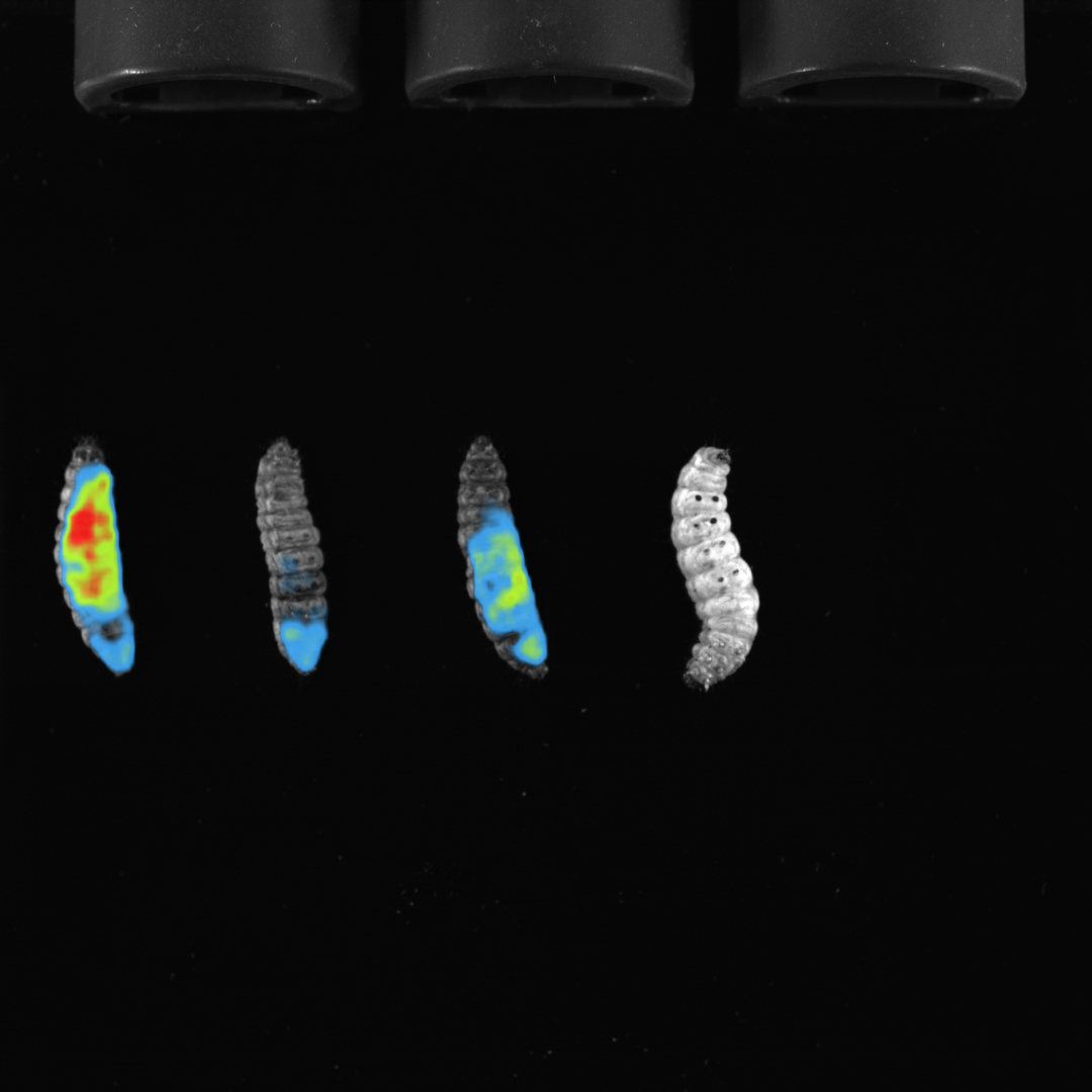
Newton 7.0 Image Gallery
Applications
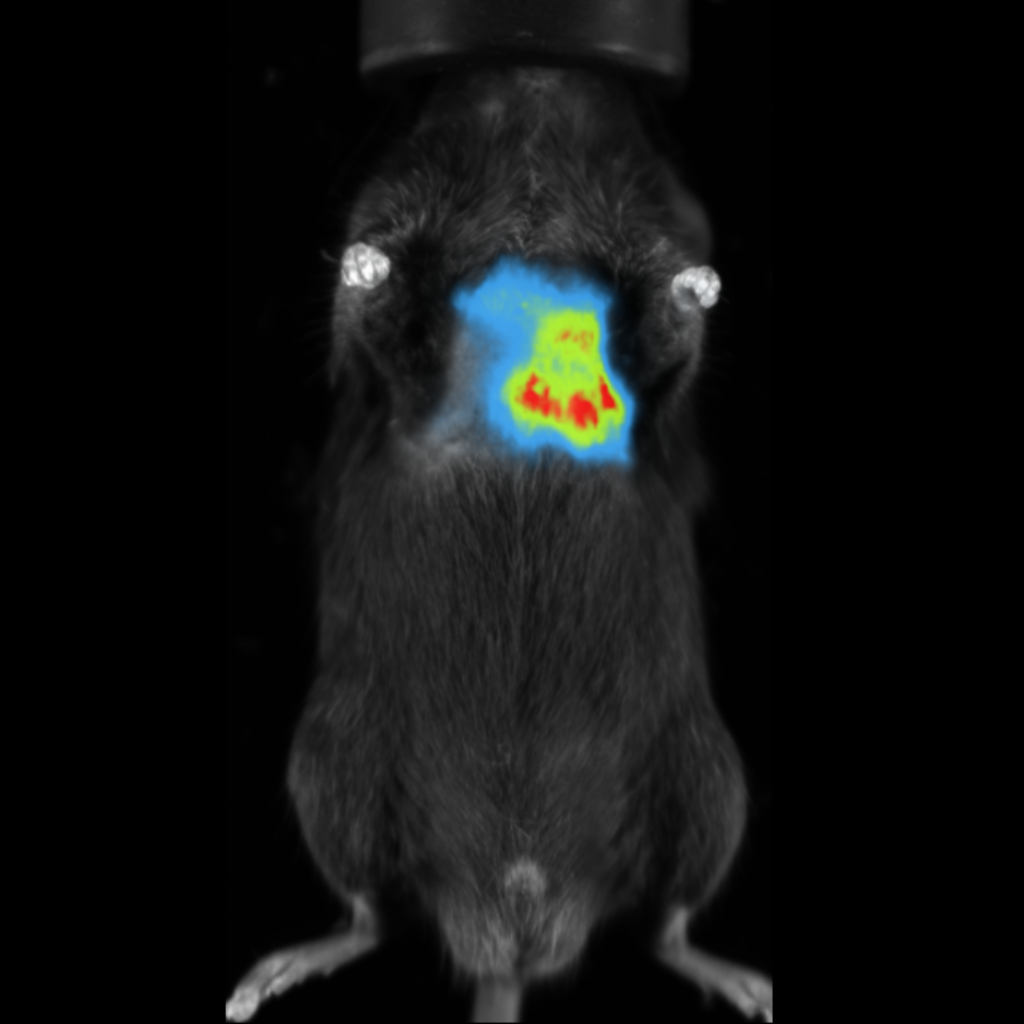
Oncology
Optical imaging can be used to non-invasively monitor the progression and spread of cancer throughout the body in preclinical animal models
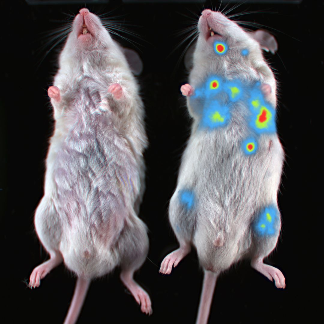
Immunology
Monitoring various populations of immune cells can contribute significantly to the understanding of their physiology and the development of new therapeutic strategies
Infectious disease
Optical imaging can be used to non-invasively visualize a site of infection as well as the efficacy of a treatment in the context of living subject
Neurology
Optical imaging can be used to monitor the progression of various neurodegenerative diseases as well as to test novel targeted therapeutics within the brain and spinal cord
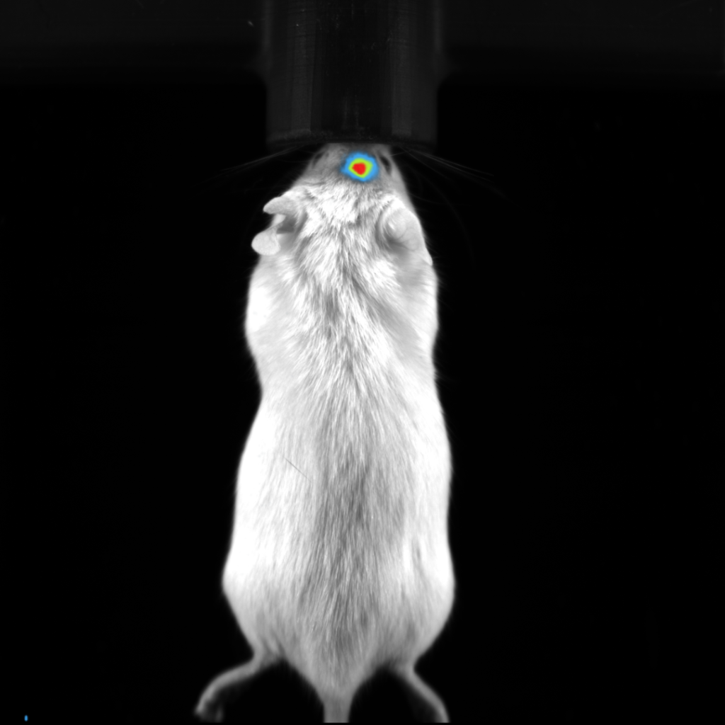
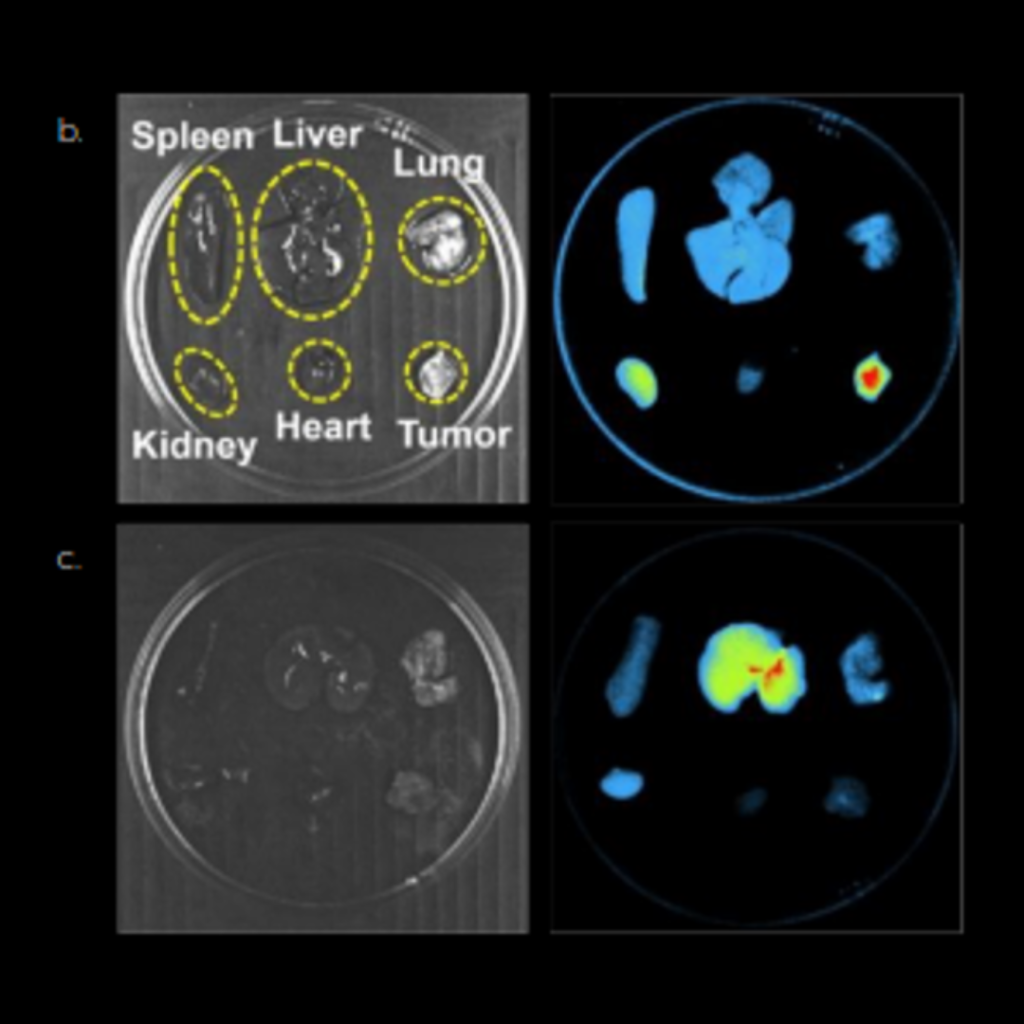
Biodistribution studies
The ability to image the whole subject, gives optical imaging a unique advantage in preclinical biodistribution studies, one image can provide measurements for multiple organs throughout the body
Publications & Articles
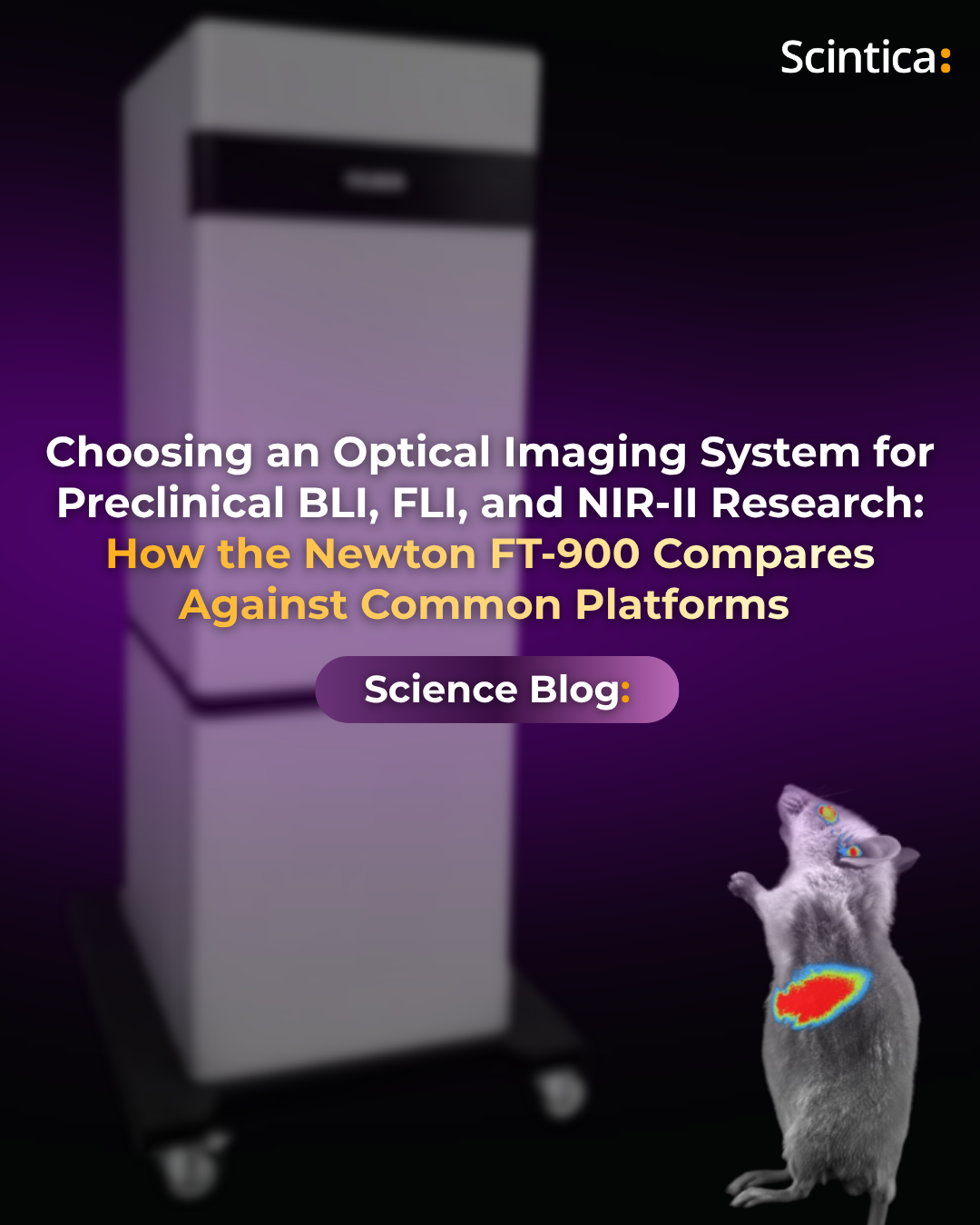
Choosing an Optical Imaging System for Preclinical BLI, FLI, and NIR-II Research
Choosing an Optical Imaging System for Preclinical BLI, FLI, and NIR-II Research: How the Newton FT-900 Compares Against Common Platforms
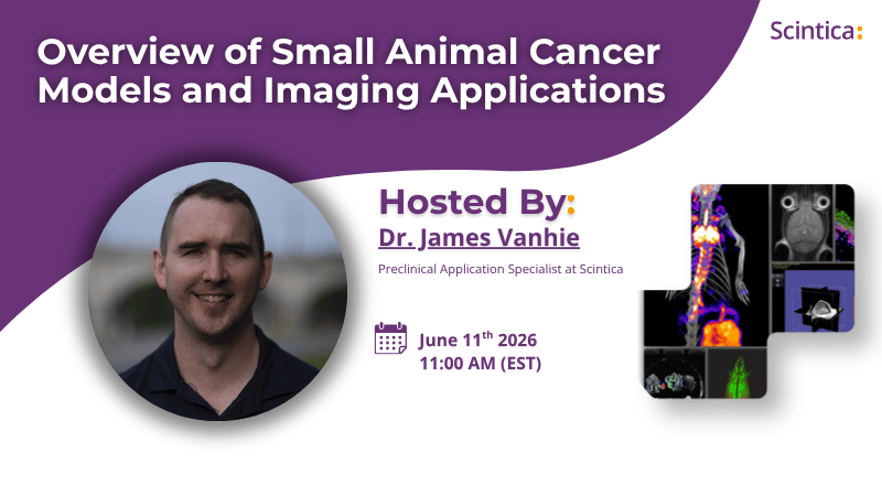
(June 11th, 2026) Webinar: Overview of Small Animal Cancer Models and Imaging Applications
(June 11th, 2026) Webinar: Overview of Small Animal Cancer Models and Imaging Applications In this webinar, we reviewed common preclinical
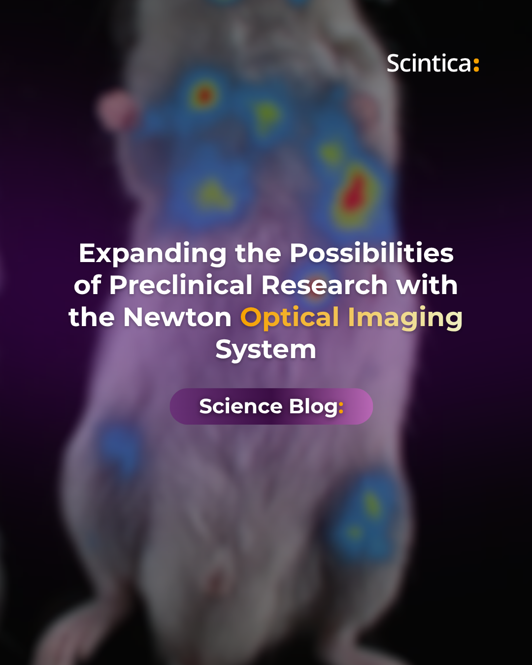
Expanding the Possibilities of Preclinical Research with the Newton Optical Imaging System
Expanding the Possibilities of Preclinical Research with the Newton Optical Imaging System Understanding biological processes as they occur in living
