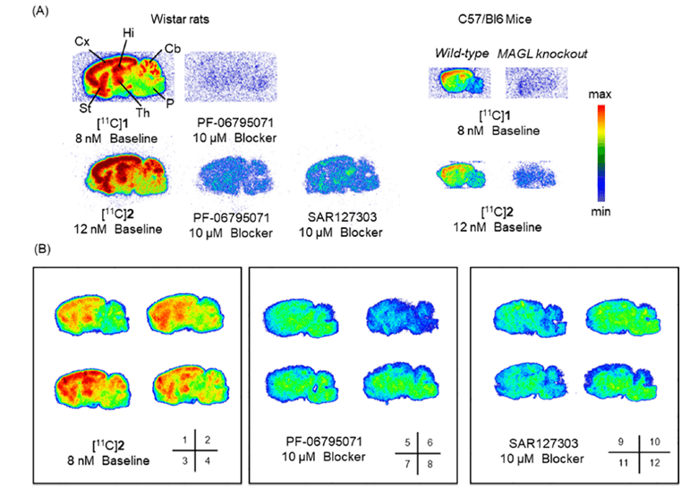
Discovery, Synthesis, and the Evaluation of a Novel PET Tracer for Monoacylglycerol (MAGL): A Short Article Review
System Used:SuperArgus PET/CT Download PDF Here The endocannabinoid system (ECS) is not a
Preclinical Imaging of Conscious Unrestrained Animals
Preclinical Imaging of Conscious Animals System Used:SuperArgus Download PDF Here Introduction Molecular imaging
Development of [¹⁸F]FPy-WL12 as a PD-L1 Specific PET Imaging Peptide
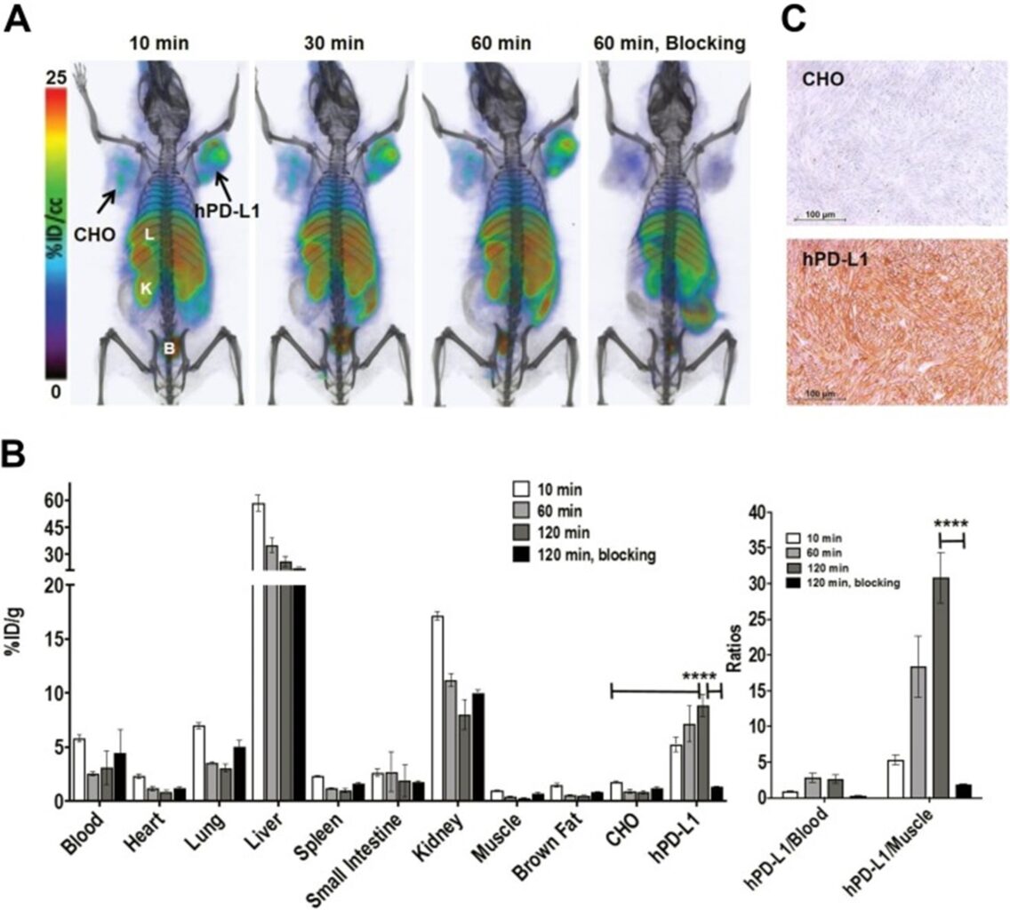
System Used:SuperArgus Programmed death ligand 1 (PD-L1) is the principal ligand of