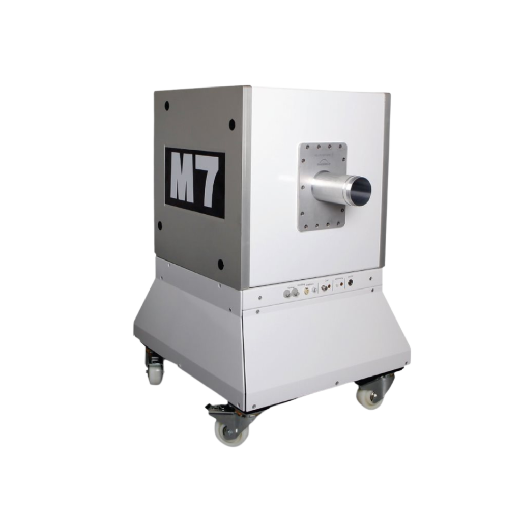
M-Series Compact MRI
High Performance Preclinical Magnetic Resonance Imaging (MRI)


System Overview
The M-Series™ systems are cryogen/cooling-free, self-shielded, high-performance MRI systems based on permanent magnet technology. The M-Series systems allow preclinical researchers, with or without in-depth knowledge of MR physics, to utilize the gold standard method in soft tissue imaging without the cost, complexity, and technical burden of superconducting MRI systems. Varying-sized models allow for imaging of mice, rats, similarly sized species/samples, as well as brains of non-human primates. Based on the permanent magnet technology, there are no special infrastructure requirements, operating and maintenance costs are minimal, and they can be easily integrated into existing lab space. These 1 Tesla systems are optimized for anatomical, functional, and molecular imaging applications in cancer, cardiac, neuroscience, and multimodal imaging. The optional SimPET insert can be integrated with the M-Series for simultaneous PET/MRI acquisition.
There are no special infrastructure requirements, operating and maintenance costs are minimal, and they can be easily integrated into existing lab space. These 1 Tesla systems are optimized for anatomical, functional, and molecular imaging applications in cancer, cardiac, neuroscience, and multimodal imaging.
The animal handling system ensures the stability of the animal throughout the imaging session with easy-to-use fully integrated anesthesia, heating, and physiological monitoring systems.
The optional SimPET insert can be integrated with the M-Series for simultaneous PET/MRI acquisition.
MR contrast agents further enhance the capabilities of this imaging modality, providing invaluable insights into tissue/tumor perfusion, myocardial infarction size, and molecular targets. Gadolinium and Iron-Oxide based contrast agents can be used with the M-Series product line. These systems are ideal for contrast agent imaging as it operates at 1 Tesla (T), which embodies an increased sensitivity to the effects of Gadolinium compared to higher-field MR systems.
Features & Benefits
The cost effective and compact M-Series magnetic resonance imaging (MRI) systems generate high-resolution 3D whole body, anatomical, functional and molecular images in small animals
Self-shielded permanent magnet
The M-Series systems are permanent magnets, as such they can be installed within an existing animal facility or laboratory space next to other equipment or furnishings.
The citing options are endless and costs to modify an existing space are minimal:
- System is self-shielded, so no faraday cage or extra shielding is needed to protect the magnet from the surroundings or the surroundings from the magnet.
- Permanent magnets do not generate heat to create the magnetic field, so no liquid (helium or water) cooling is needed, along with the additional equipment and space required for this
- Only a single 220V plug is required, electricity is only required during imaging to power the fans, gradients, and other electronics.
Positioning of the system components (magnet and electronics) are quite flexible and can be up to 7 meters from one another.
Affordable purchase price for fully-equipped systems
MRI, the gold standard in soft tissue imaging, is more accessible for preclinical researchers.
Fully integrated physiological monitoring, heating, & anesthesia
Coils are self-tuned and matched, and the system performs calibrations automatically.
The M-Series systems provide a simplified workflow from animal preparation to image acquisition, without the need for time consuming system calibration and set-up steps to ensure optimization of the imaging subject with the coil and the rest of the system.
Real-time monitoring of the imaging subject ensures stability of the animal throughout the imaging session.
Image acquisition may be triggered off of either the respiratory or cardiac signals.
No electricity is required to maintain the magnetic field, so no heat dissipation (cryogens or water) is needed
As the M-Series systems utilize a permanent magnet the operating and maintenance costs are minimal:
- No constant supply for electricity is required to maintain the magnetic field
- No heat is generated to create the magnetic field, so no additional equipment or maintenance of that equipment is required (i.e. helium or water pumps and compressors)
- There are minimal maintenance requirements on the M-Series. Simply a yearly preventative maintenance visit, but no daily, weekly, or monthly requirements by the user.
User-friendly software interface with numerous sequences
Preclinical researchers, without prior knowledge of MR physics, are able to utilize the optimized default sequences for some of the most common imaging applications. This allows high quality images to be reproducibly obtained of their specific animal models using the M-Series systems.
Minimal animal preparation steps
Imaging subjects must be still during acquisition, so animals should be lightly anesthetized. This can be done using the integrated anesthesia system. Once ready the imaging subject is placed into the animal handling platform and positioned within the coil.
No other preparation steps are required for standard imaging, making it possible to image a cohort of animals quickly, with no need for further manipulation of the imaging subject.
Hardware and software add-ons are available to allow for integration of other imaging modalities such as PET and bioluminescence
Multimodal imaging allow to combine the exquisite anatomical information of MRI with the sensitivity and specificity of Molecular and Metabolic imaging.
Models & Specifications

M3
Mice Only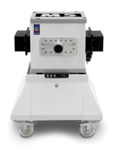
M5
Mice, Rats & Other Small Animals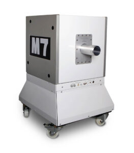
M7
Mice, Rats & Other Large Animals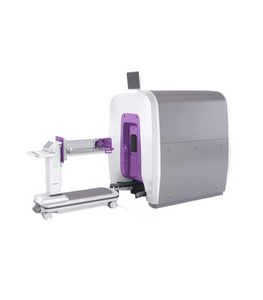
M12
Non-human PrimatesM-Series Preclinical MRI Specifications
|
Specifications Front End (Magnet Stand) |
M3 |
M5 |
M7 |
M12 |
|---|---|---|---|---|
|
Dimensions (Height x Width x Depth) |
1080 x 734 x 734 mm 42.5 x 29 x 29 inches |
1133 X 800 x 800 mm 44.6 x 31.5 x 31.5 inches |
1320 x 790 x 950 mm 52 x 31 x 37.5 inches |
1810 x 1450 x 1710 mm 71.26 x 57.09 x 67.32 inches |
|
Weight |
650 Kg / 1,430 lbs |
950 Kg / 2,095 lbs |
1,550 Kg / 3,415 lbs |
5,500kg / 12,125lbs |
|
Magnet Opening Flange Insertion Dimeter Inner Bore (Height x Width) |
70 mm / 2.8 inches 50 x 130 mm / 2 x 5.1 inches |
No insertion flange in M5 (the bore is open) 76 x 200 mm / 3 x 7.9 inches |
97 mm / 3.8 inches 220 x 90 mm / 8.6 x 3.5 inches |
184 x 260 mm / 7.2 x 10.2 inches |
|
Imaging Volume |
80 x 80 x 35 mm3 spheroid |
90 x 90 x 60 mm3 spheroid |
120 x 120 x 70 mm3 spheroid |
120 x 130 x 130 mm3 ellipsoid |
|
Bo (Tesla) |
1T |
1T |
1T |
1T |
|
Back End Electronics & Cabinet |
M3 |
M5 |
M7 |
M12 |
|---|---|---|---|---|
|
Dimensions |
1760 x 600 x 1150 mm 69.3 x 23.6 x 45.3 inches |
1760 x 600 x 1150 mm 69.3 x 23.6 x 45.3 inches |
1760 x 600 x 1150 mm 69.3 x 23.6 x 45.3 inches |
1760 x 600 x 1150 mm 69.3 x 23.6 x 45.3 inches |
|
Weight |
300Kg / 660 lbs |
300Kg / 660 lbs |
300Kg / 660 lbs |
300Kg / 660 lbs |
| Gradients | M3 | M5 | M7 | M12 |
|---|---|---|---|---|
|
Strength |
440 (X, Y), 540 (Z) mT/m |
430 (X, Y, Z) mT/m |
150 (X, Y, Z) mT/m |
150 (X, Y, Z) mT/m |
|
Slew rate (T/m/s) |
1750 T/m/s |
1720 T/m/s |
500 T/m/s |
500 T/m/s |
| All Models | |
|---|---|
|
|
20-240 VAC, 15A, single phase in countries using 220-240V
208 VAC, 15A, single phase in countries using 120V |
| Pulse Sequences | Procedures |
|---|---|
|
Procedures are pulse sequences that are combined with dedicated post-processing routines.
|
Animal Handling System
Fully integrated animal handling and coil system includes:
- Beds that suite a variety of sized animals such as mouse, rat, and non-human primate
- Anatomy specific coils to enhance signal to noise ration and image quality
- Fully integrated anesthesia delivery and gas exhaust scavenging
- Individually controlled, multi-point, isoflurane vaporizer
- Continuous monitoring of ECG, heart rate, respiration rate, and body temperature
- Allows for ECG and/or respiratory triggering
- Heated water is continuously circulated to maintain body temperature
RF Coils
| Type | Inner Diameter | Lenght |
|---|---|---|
|
Mouse Head |
23 mm |
25 mm |
|
Mouse Body |
30 mm |
50 mm |
|
Mouse Whole Body |
30 mm |
80 mm |
|
Rat Head |
35 mm |
40 mm |
|
Rat Body |
50/60 mm ellipsoid |
90 mm |
|
Large Rat Body |
71 mm |
90 mm |
|
Non-human Primate |
143 mm |
120 mm |
RF Coils
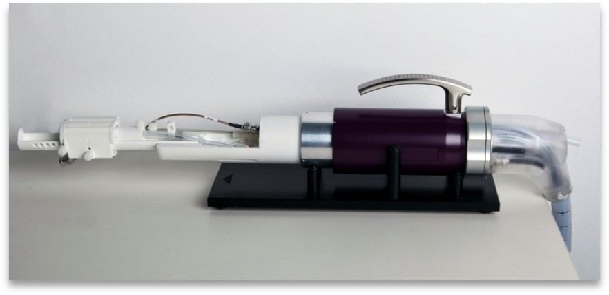
Accessories & Add-ons
The SimPET insert is a state-of-the-art, compact, insert with low power consumption and excellent PET detector stability that fits within the M7 MRI. The SimPET is based on advanced silicon photomultiplier (SiPM) technology for truly simultaneous PET/MR imaging, affording combined sensitive molecular and metabolic information with the high-resolution anatomical soft-tissue context for a wide range of applications in neurology, cardiovascular biology and oncology.
M-Series Imaging Gallery

Anatomy & Morphology of mouse abdomen: T1- and T2- weighted scans of wildtype mouse body abdomen
Neurobiology of mouse brain: T2- weighted images of a mouse brain
Epilepsy in the rat brain: T2- weighted images of a rat brain 48 hours after induction of epilepsy
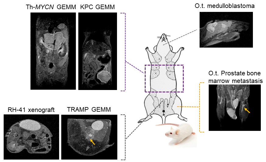
Tumor visualization in various cancer models: Fat-suppressed T2-weighted imaging can be used to detect and quantitatively characterize the growth of a wide range of cancer models
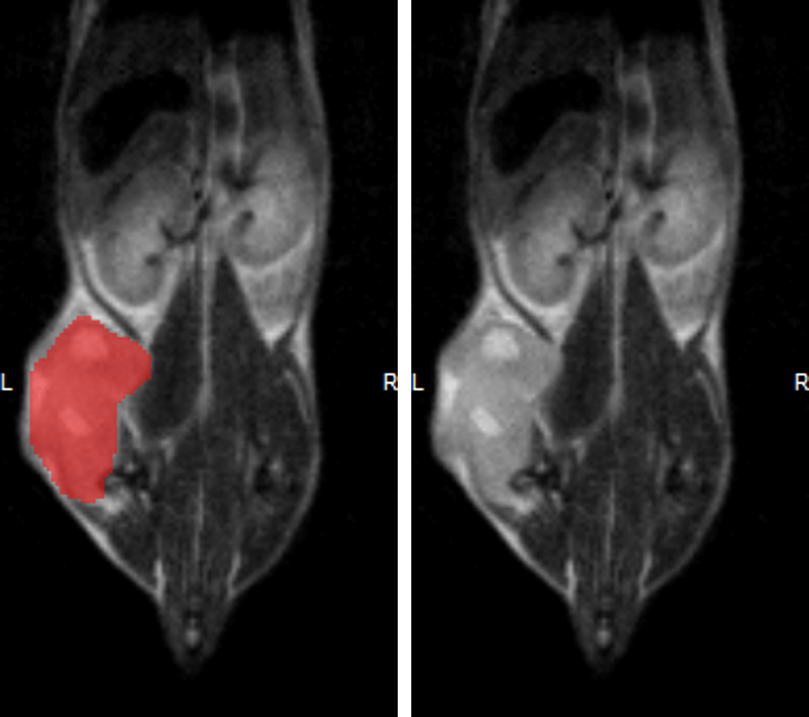
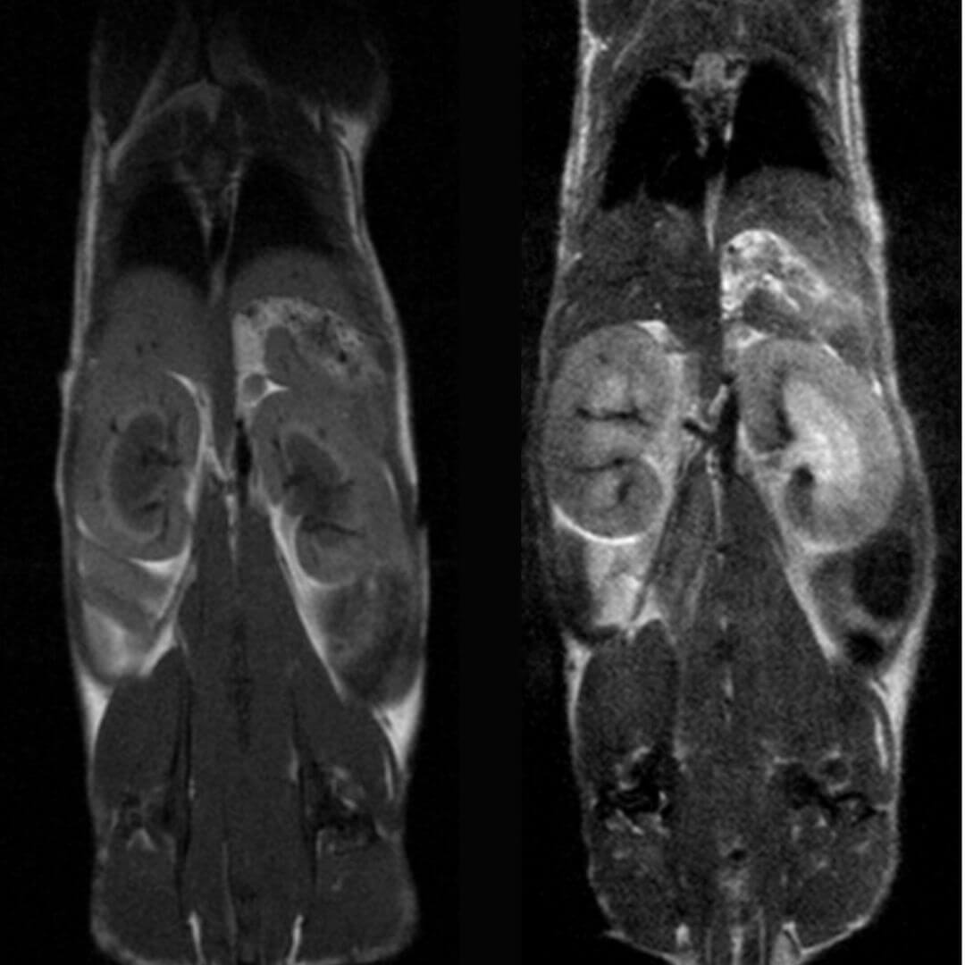
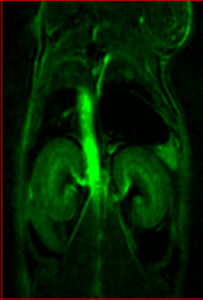
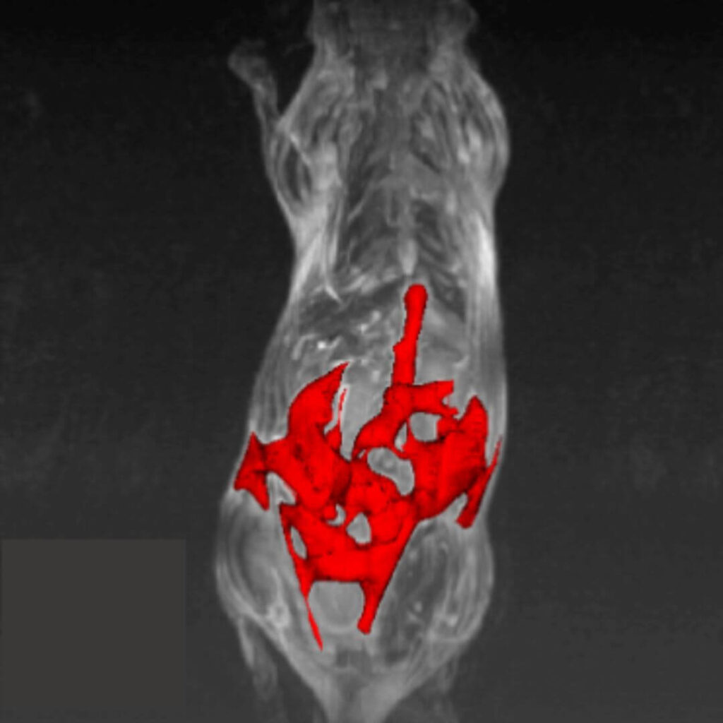
Hindlimb tumor growth: Monitoring the growth of xenograft tumor grown in the mouse hindlimb is identified with T2-weighted images. Segmentation of tumor region of interests (in red) on each tumor-containing slice allows accurate volume quantification
Cardiac Cine loop images: Long axis view of a single slice of the heart was acquired with Cine loop images. Long axis images can be used for strain analysis in third party softwares
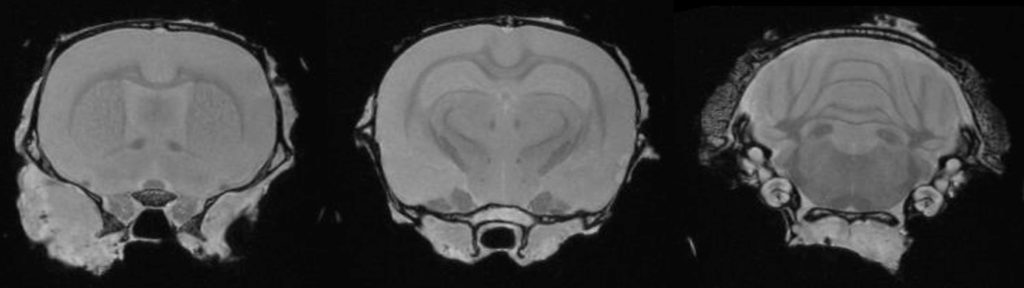
Ex Vivo Imaging: Ex vivo imaging can produce high-resolution images of the rat brain providing excellent delineation of brain regions

Multimodal Imaging: Multimodal imaging combines the strength of MRI with other imaging modalities such as PET and CT. PET provides information on the spatial distribution of molecular signal of interest. In this case MRI confirmed that the absence of tracer uptake in the center of the images was due to the presence of a necrotic core, which appears hyperintense on T2-weighted MRI
Applications
Anatomy & Morphology
The M-Series systems can be used to study a wide variety of anatomical and morphological imaging targets including:
- Inflammation
- Metabolic disorders, including diabetes and obesity
- Specific organ pathology, i.e. liver, kidney, spleen, etc.
- Tissue perfusion with contrast agent
T1- and T2- weighted images of Wild-type mouse abdomen
Maximum intensity projection show visceral fat segmentation from T1-weighted images of mouse abdomen
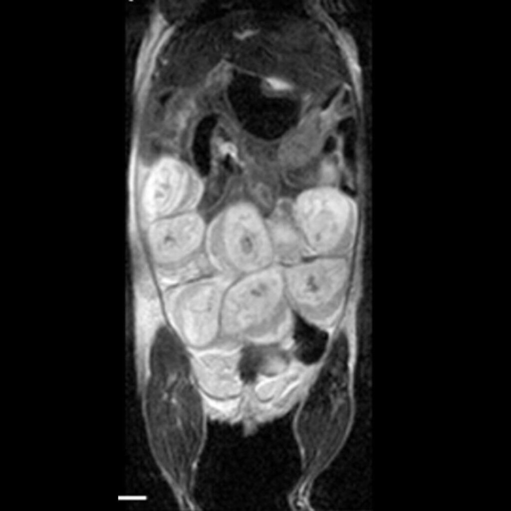
T2-weighted images show mouse placenta and fetal development during pregnancy
Neurobiology
The M-Series systems can be used to study a wide variety of neurological diseases and disorders including:
- Inflammation
- Stroke
- Epilepsy
- Neurodegeneration
- Tumor biology
- Anatomy
- Cerebral perfusion with contrast-enhanced angiography
- Targeted molecular imaging with contrast agents
- Traumatic Brain Injury (TBI)
T2- weighted images of a mouse brain.
T2- weighted images of a rat brain 48 hours after induction of epilepsy.

T2- (top row) and T1- (bottom row) weighted images show stroke lesion and its segmentation (in red) within the rat brain.
Cancer/Oncology
The M-Series systems can be used to study a variety of tumor models, and stages of disease. As no manipulation of the tumor cells are required models including xenografts, orthotopic, transgenic, and patient-derived xenografts can be imaged in a wide variety of imaging subjects to focus on:
- Tumor detection
- Quantitative monitoring of tumor progression
- Quantitative response assessment for drug studies
- Tumor phenotyping including necrosis detection
- Functional imaging of tumor hemodynamic micro-vasculature and hypoxia (w/o contrast agents)
- Targeted molecular imaging with contrast agents
Fat-suppressed T2-weighted imaging can be used to detect and quantitatively characterize the growth of a wide range of cancer models.
Monitoring the growth of xenograft tumor grown in the mouse hindlimb is identified with T2-weighted images. Segmentation of tumor region of interests (in red) on each tumor-containing slice allows accurate volume quantification.
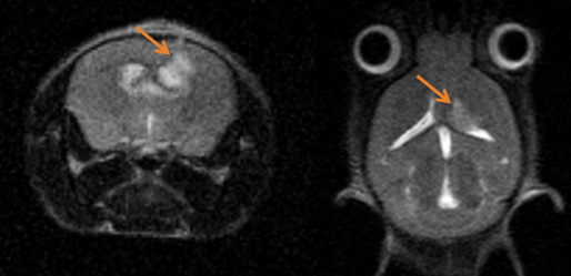
Monitoring tumor growth in the mouse brain, following the orthotopic injection of glioblastoma cells, using T2-weighted images.
Cardiovascular Biology
The M-Series systems allow for cardiac images to be acquired from a wide variety of imaging subject to assess the following:
- Long and short axis acquisition
- Systolic/Diastolic Volum
- Ejection fraction
- Fractional shortening
- Wall thickness
- Strain and torsion
- Myocardial perfusion
- Angiography with contrast agents
All parts of the heart and surrounding vasculature can be easily imaged including the left and right atrium which are otherwise quite difficult to image by other imaging modalities.
Slice prescription to acquire reproducible images is also simplified on the M-Series system, allowing for reproducible data over the course of a longitudinal study.
Fat-suppressed T2-weighted imaging can be used to detect and quantitatively characterize the growth of a wide range of cancer models.
Short axis view of a single slice of the heart was acquired with Cine loop images. Short axis images can be used for analysis of functional measurements in third party softwares.
Ex vivo Imaging
MRI can be performed on excise tissues fixed in formalin at resolution way higher than in a live animal. Ex Vivo 3D MRI affords the detection and quantification of lesions in a whole organs or structure within in the brain and can help guiding further histopathological processing and sectioning for conventional pathological examination of regions of interest.
- High-resolution imaging
- Lesion detection
- Volume quantification
Ex vivo imaging can produce high-resolution images of the rat brain providing excellent delineation of brain regions.
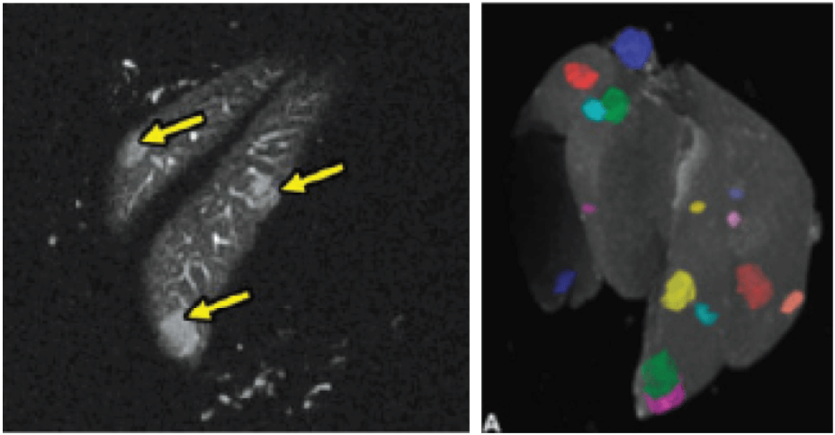
Ex vivo imaging of liver, allowing lesions to be identified, counted and measured (volume) as well as guide tissue sectioning for conventional pathological examination.
Multimodal Imaging
Current trends in preclinical imaging see multimodal imaging as a means of obtaining synergistic information about a specific disease model, or response to target compound, and should be, when possible, considered in all studies.
The M-Series allows for integration with the SimPET insert, for simultaneous PET/MRI.
Alternatively, a multimodal imaging cassette has been designed to fit within the MRI coil on the M-Series, allowing for transportation of the animal to other imaging modalities. Co-registration of these images may be done in third party software such as VivoQuant.
Multimodal imaging combines the strength of MRI with other imaging modalities such as PET and CT. PET provides information on the spatial distribution of molecular signal of interest. In this case MRI confirmed that the absence of tracer uptake in the center of the images was due to the presence of a necrotic core, which appears hyperintense on T2-weighted MR
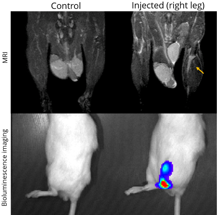
MRI is a complementary method to bioluminescence imaging in the detection of tumors allowing more precise assessment of the location and extent of orthotopic tumors (here an orthotopic model of prostate bone marrow metastasis).
Contrast Agent-Enabled Functional and Molecular Imaging
As the M-Series systems all operate at 1T, they are well suited for use with T1 contrast agents such as Gadolinium (Gd) or Manganese (Mn). There contrast agents provide a higher signal enhancement at lower field strengths, such as 1T, when compared to higher field strengths such as 3T, 7T or higher.
Additionally, T2 contrast agents can be used with the system, allowing for full flexibility.
There are a wide variety of both functional and molecular imaging capabilities enabled by contrast agents:
- Targeted molecular imaging
- Stem cell tracking
- Perfusion and vascular imaging
- Dynamic Contrast Enhanced (DCE) acquisitions
- Delayed contrast enhanced imaging in the heart to look at infarct size and tissue viability in the transmural region
MR angiography with high T1 relaxivity liposomal-Gd contrast agents produces reconstructed 3D map of the cerebral vasculature of a mouse brain.

Dynamic contrast -enhanced (DCE-) MRI shows the change in T1-signal following the remote intravenous injection of a T1 contrast agent via the lateral tail vein, which can be used to quantify vascular perfusion.
Signal enhancement maps generated by subtraction of the pre-contrast T1-weighted images from post contrast images using third party software VivoQuant.