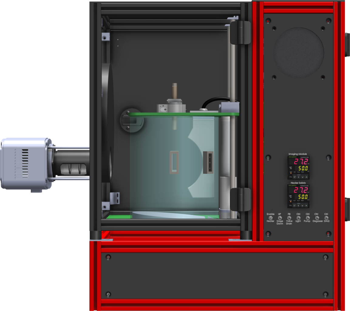
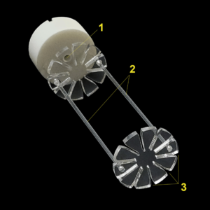
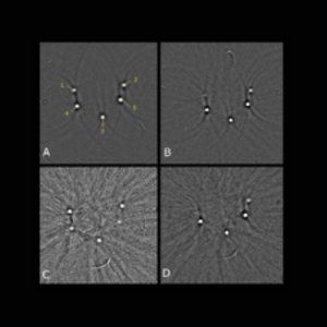
Microcuvette Sample Holder
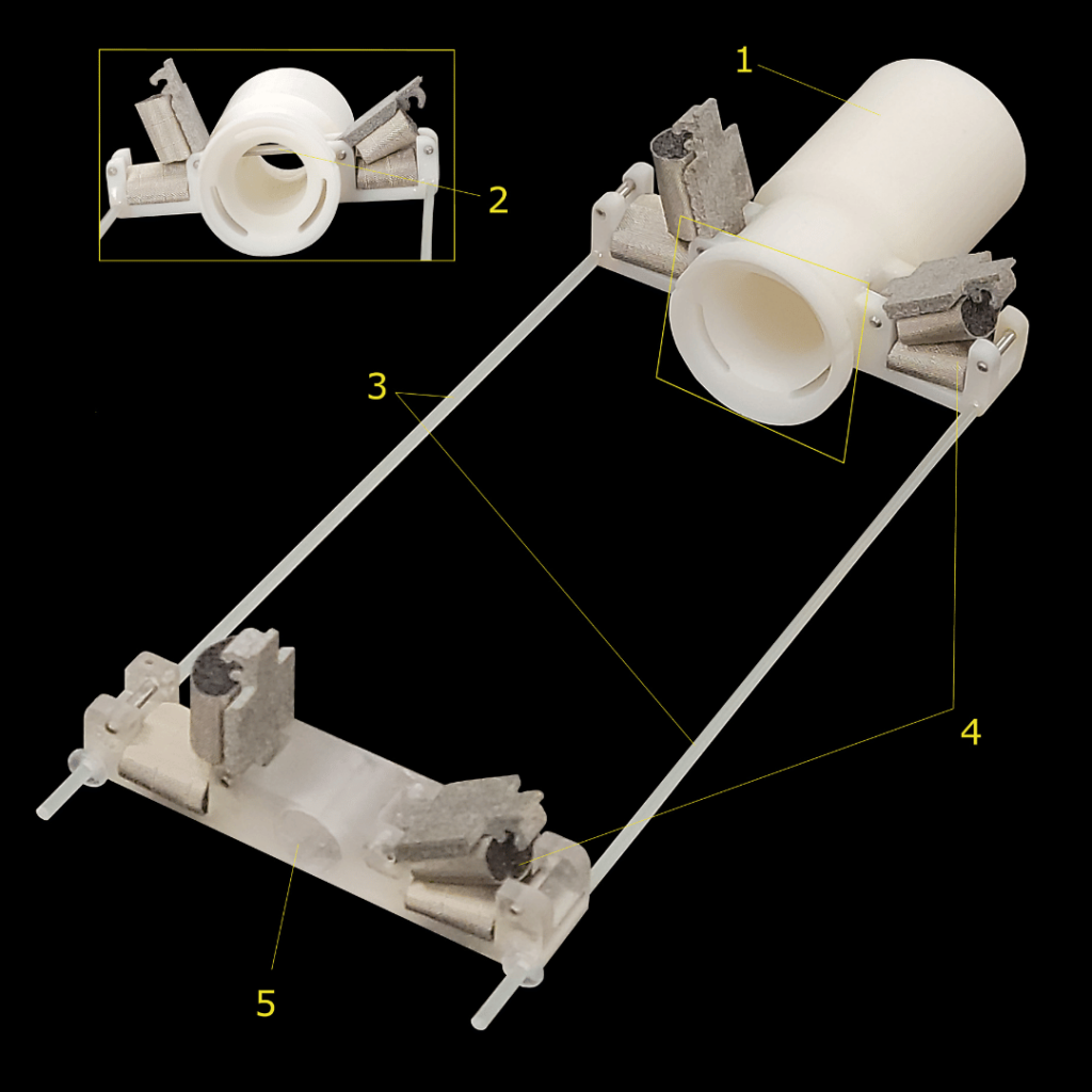
Mouse Holder
Microcuvette Sample Holder
Mouse Holder
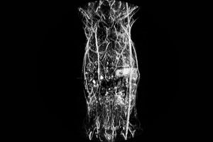
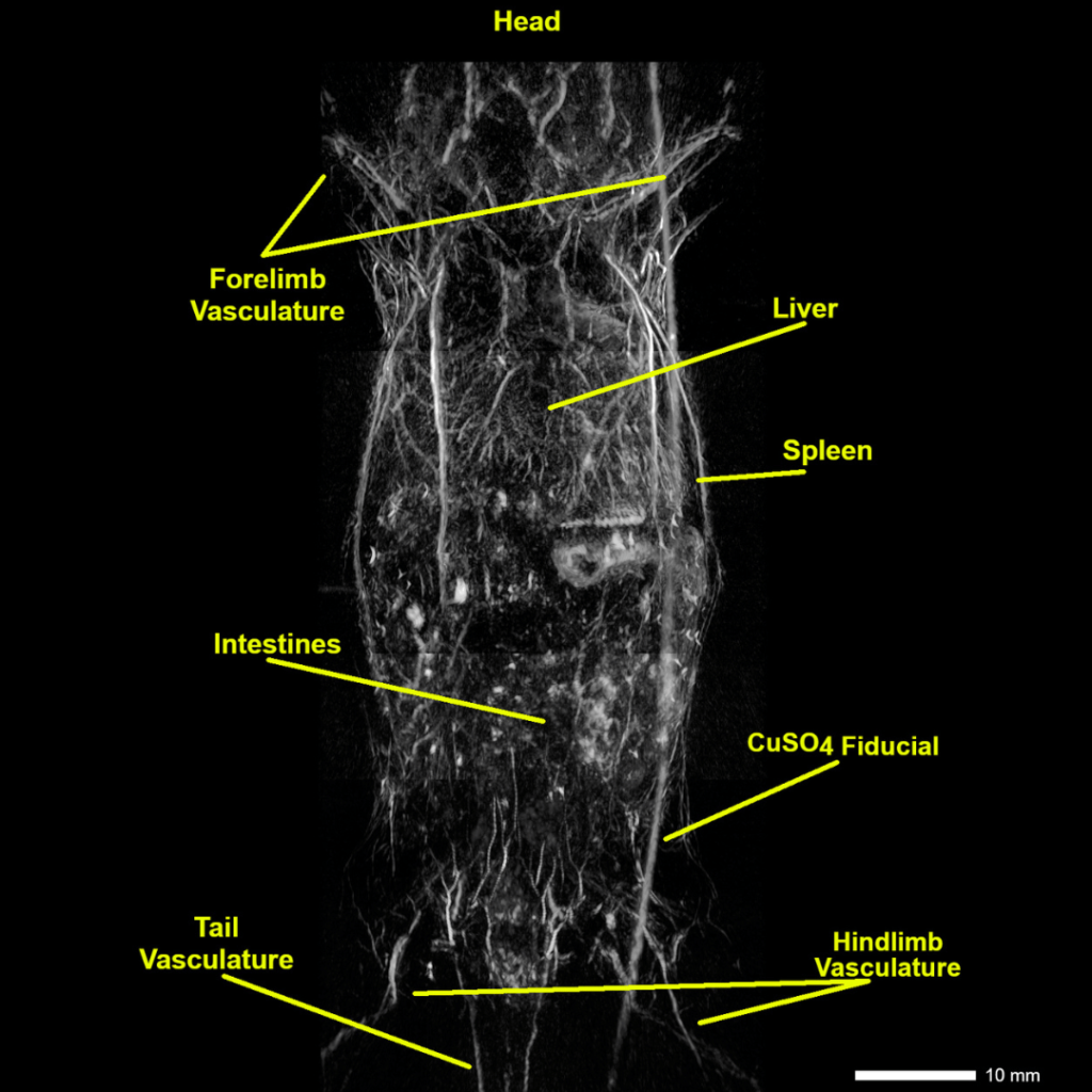
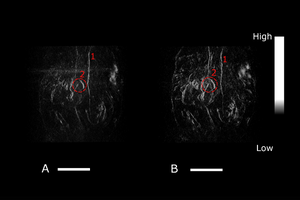
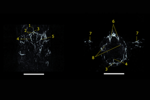
Anatomical Imaging / Registration
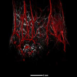
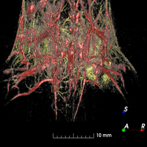
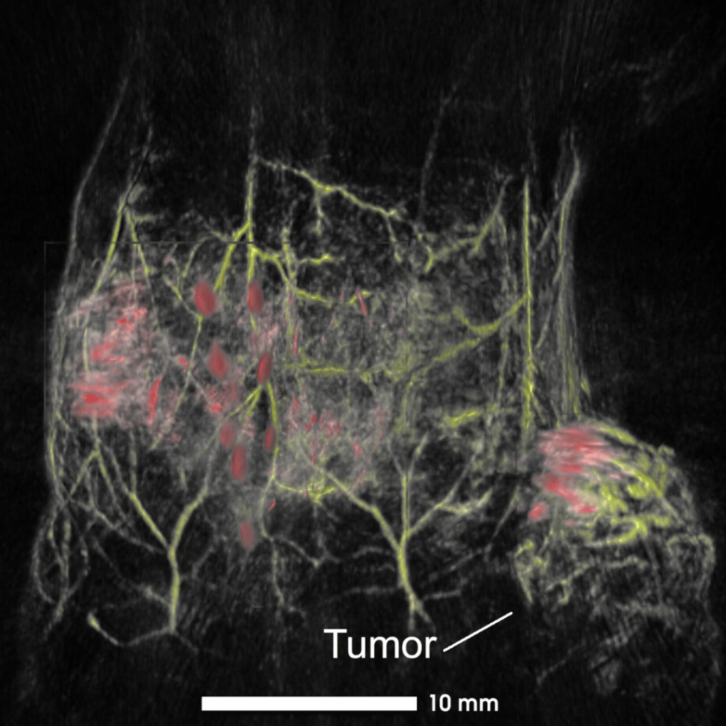
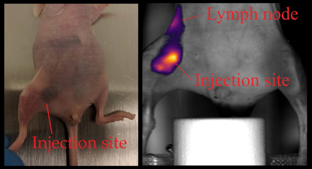
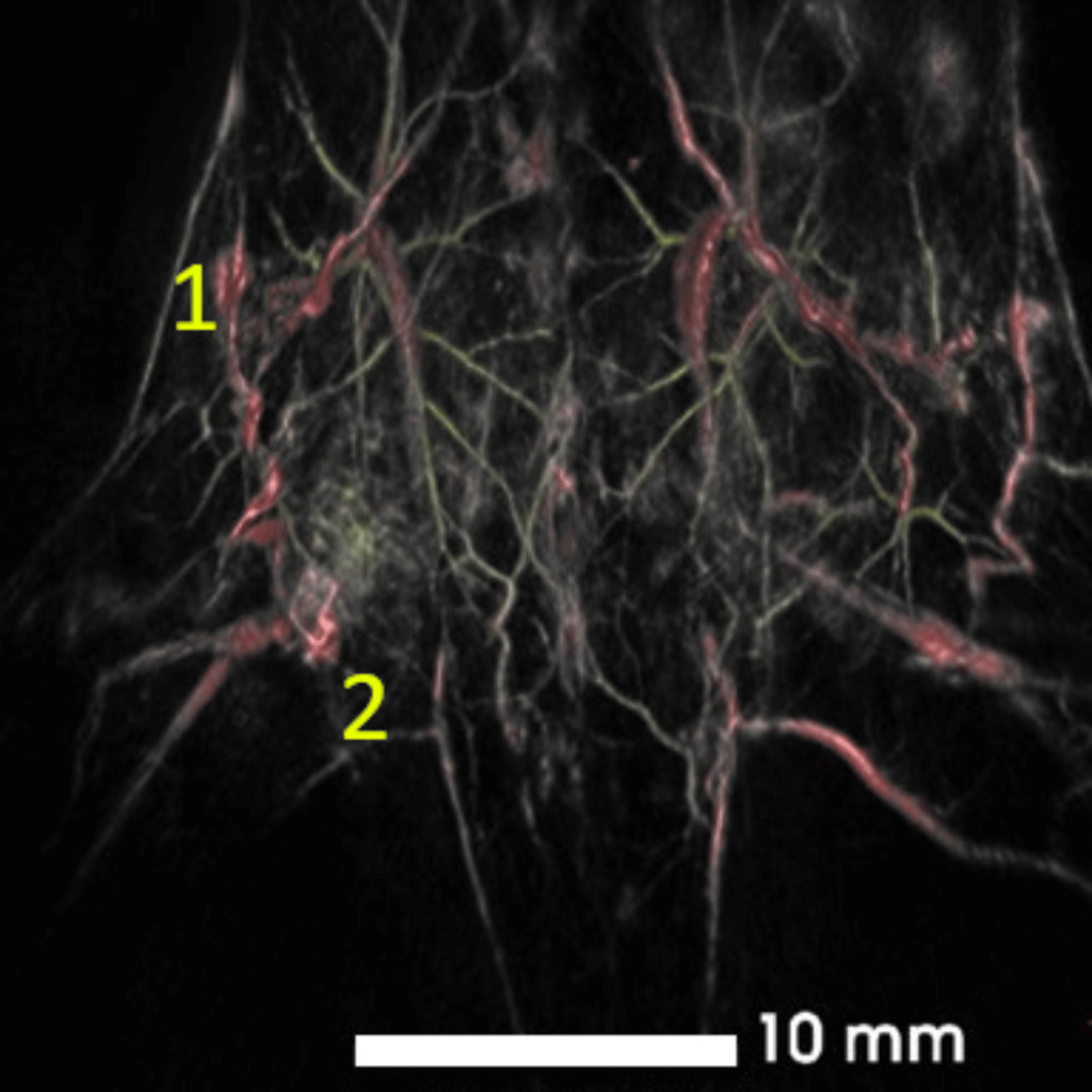
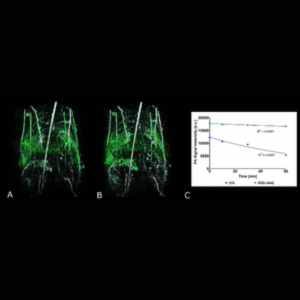
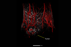
Cancer
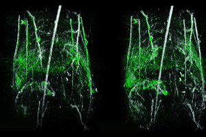
Tissue Engineering & Regeneration
Developmental Biology
Neuroimaging
Molecular probes, optical contrast agents, and fluorophores
Anatomical Imaging / Registration
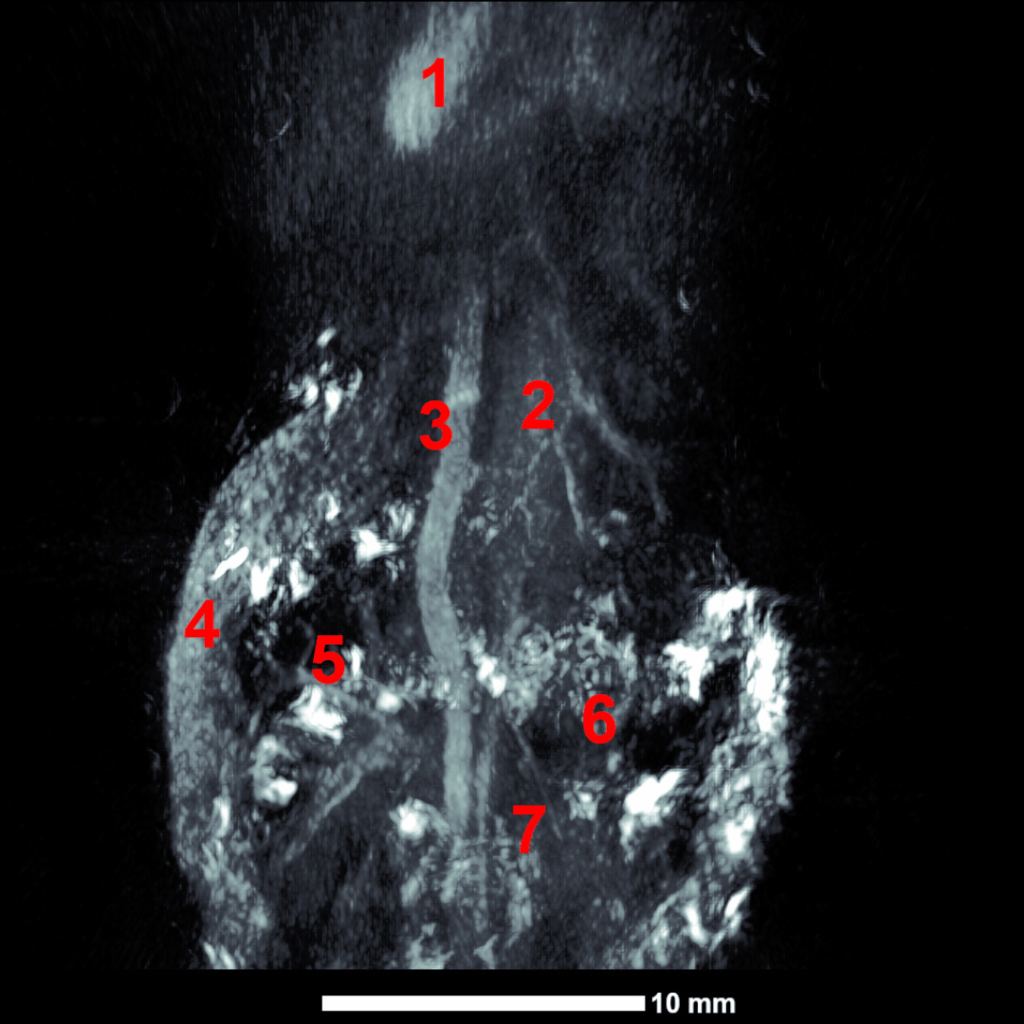
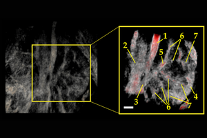
Cancer
Tissue Engineering & Regeneration
Developmental Biology
Neuroimaging

Molecular probes, optical contrast agents, and fluorophores