Gel Count TM
The "gold standard" colony and spheroid counter for cancer researchers

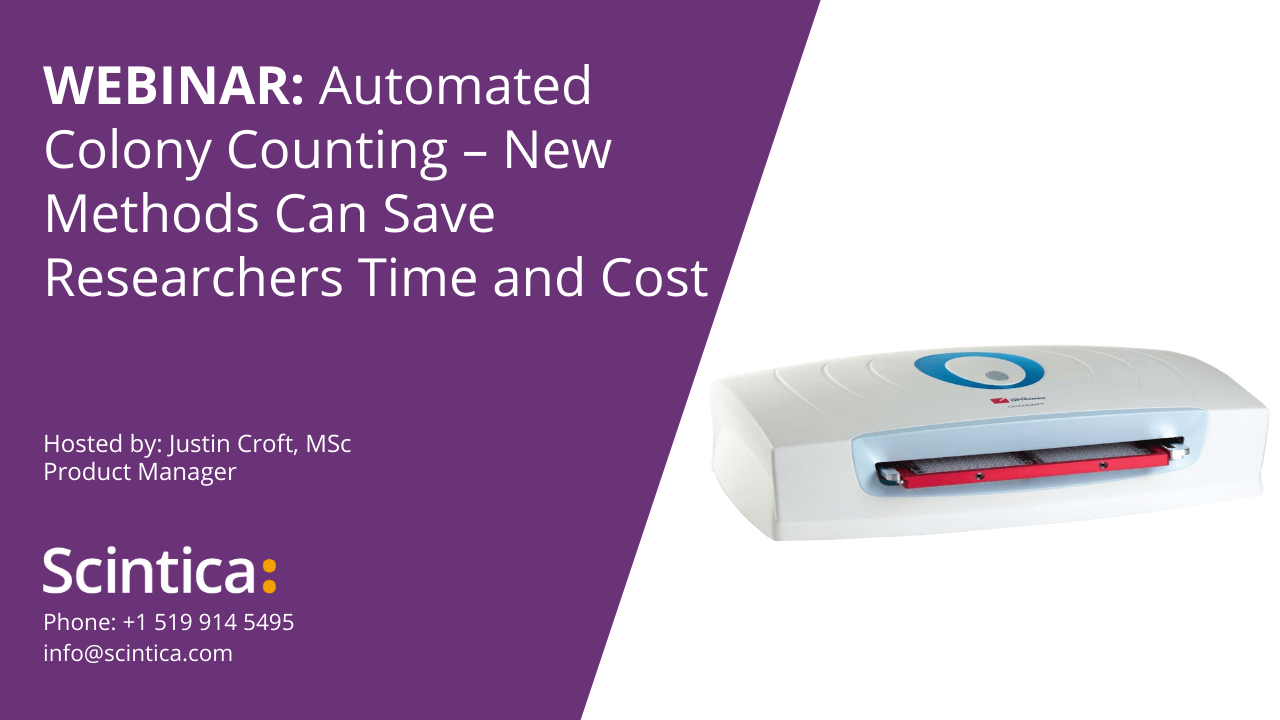
Webinars
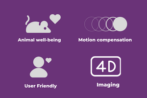
Features
System Overview
The GelCount is known as the “gold standard” for cancer biologists performing colony counts and is backed up by over 200 scientifically peer reviewed articles.
The Colony Formation Assay is universally recognized as an accepted method for measuring the effects of cell viability when exposed to chemotherapy drugs, radiation or other agents. However, manual counting the subsequent cell colonies is a difficult and painstaking task in which objectivity is often tough to achieve.

Introducing GelCount™ 2, an integrated, PC software-operated imager and analysis platform that automates the detection, counting and analysis of 3D mammalian cell colonies, organoids or spheroids in multi-well plates and Petri dishes.
With over 500 scientific citations to its name the original GelCount™ has become a benchmark for biologists around the globe. GelCount™ 2 introduces a revised design, faster
processing, and a more intuitive user experience.
Features & Benefits
A fully integrated colony counter
The GelCount is the total-package solution for imaging, counting and characterizing adherent (2D) or non-adherent (3D) colonies with a combined hardware and patented PC software program. Colonies are fully imaged and are saved to a computer where they can be processed and characterized. The data generated can easily be exported from this single, integrated platform. As a result, the GelCount has removed the unsatisfactory alternative of imaging colonies on one device, then relocating these images to and processing them on separate image analysis software.
Image output & exporting data
Sample images can be stored in one of two ways. First, colony images can be saved in a raw format for a permanent archive of colony samples and ‘off-line’ processing or re-processing on any workstation with the free GelCount software. Secondly, these images can also be stored in a generic bitmap format for publication or presentation quality images. All colony counts, and other statistical data are automatically exported to Microsoft Excel.
Colony size data
In addition to yielding colony counts, the GelCount software also generates detailed colony diameter data in the form of a mean per sample, a histogram distribution, or even on an individual colony basis if necessary. This capability to quantitatively measure the effects of anti-cancer therapeutic treatments not only on total colony numbers but also on colony size greatly extends the sensitivity of colony forming assay’s by offering the user previously unavailable information on colony growth dynamics.
Diameter advantage
Not only does the GelCount generate a numerical count of
colonies, spheroids or organoids, crucially it also automatically
yields object size information (diameter in microns), providing
hitherto unavailable insight relating to cellular growth dynamics.
Workflow optimization
The software can be installed on unlimited other workstations.
Images generated by the GelCount can be stored, transferred
to and processed ‘offline’ at the user’s convenience without
tying up the imager for other users. Meanwhile colony, spheroid
or organoid counts, diameter statistics and other parameters
are exported directly to Excel®.
High throughput imaging of 3D cultures
Adherent colonies or non-adherent colonies, spheroids, and
organoids in 3D media or suspension culture can be imaged
in a single pass without z-stacking. Typical imaging and
processing times of less than 10 minutes for four 6-well plates
at typical resolution, or less than 25 minutes for four 96-well
plates at maximum resolution provides substantial throughput
gains compared to manual microscope processing.
Automated high throughput counting
The GelCount allows users to move quickly from colony samples to colony counts, plus provides extra information, like colony size data, through an easy to navigate user interface. A behind the scenes, well developed “CHARM” algorithm impartially applies user defined colony detection guidelines. These guidelines contain inclusion or omission of colonies based on colony size (diameter), distinguishing overlapping colonies, and curbing false positives from contamination and/or artefacts. Therefore, the GelCount not only significantly improves throughput, but its complete objectivity and consistency eliminate human error due to subjective interpretation, observer bias or just plain fatigue – an especially large problem when using a microscope to manually count colonies.
Versatility in use
The GelCount is appropriate for counting and analyzing both adherent monolayer colonies and non-adherent colonies in suspension (spheroids) or semi-solid matrix like methylcellulose and soft agar. Additionally, spheroids or non-adherent colonies do not normally require staining to use the GelCount.
2-year comprehensive product warranty
The GelCount comes with a full 2-year manufacturer’s warranty, covering defects in material or workmanship. An extended warranty package is available, providing up to 6 years of ‘peace of mind’ coverage, including preventative maintenance servicing which is offered at the time of purchase.
*This is resolution-dependent; mentioned example is for conventional high-resolution imaging of soft agar colonies.
** Falcon® T25 flasks (model 353082) and Sarstedt® T25 flasks (model 83.1810) are supported by the GelCount.
Objective, unbiased output
Inherent ‘machine’ objectivity and consistency eliminates
human error due to subjective object interpretation, bias or
plain fatigue – a particularly acute problem when manually
processing spheroids and organoids under a microscope.
2-year comprehensive product warranty
Our comprehensive 2-year product warranty covers defects in
material or in workmanship with optional extended warranty
and preventative servicing packages available.
On screen high-resolution view
Entire Petri dishes or wells can be conveniently observed at high resolution on-screen.
Performance
By utilizing the single pass, high depth-of-field scanning technology, linked with a simple sample-loading procedure, the GelCount delivers unmatched colony detection performance including the ability to resolve overlapping colonies and differentiate true colonies from artefacts or debris. The processing and imaging time for adherent colonies is about *6 minutes (four 6-well plates; 600 ppi) and for non-adherent colonies 12 mins (four 6-well plates; 1200 ppi). Further, the GelCount can detect colonies as small as 30 µm in diameter within gel or medium layers of up to 5 mm in depth.
Unrestricted & unlimited software licenses
The GelCount software is able to be installed on any workstation. This allows the user to save colony images on one workstation and to process them at their own convenience on another.
Flexibility and off-line processing
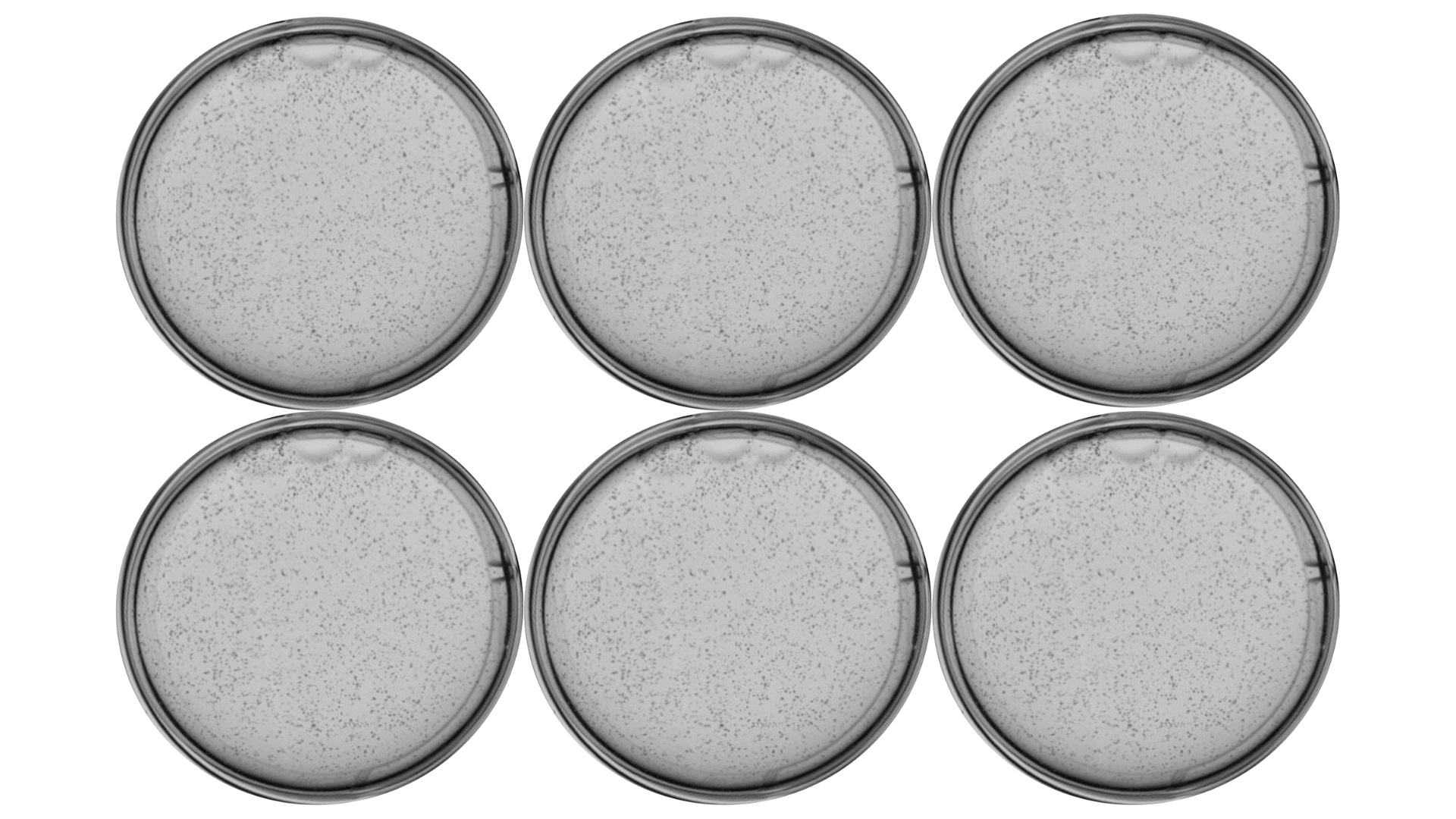
The GelCount is compatible to be used with Petri dishes (35 mm, 50/60 mm and 100 mm), multi-well plates (6-, 12-, 24-, 48- and 96-well), and some T25 flasks**. Additionally, the GelCount supports the saving of raw colony images to a PC, allowing for fully functional ‘off-line’ processing or re-processing even if the original samples are no longer available.
Free & unlimited software updates
Scintica and Oxford Optronix customer focused service includes completely free and unlimited availability of all future software updates, fixes, and enhancements for the lifetime of the GelCount.
Optional Accessories
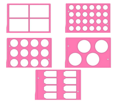
Sample loading trays
The GelCount can use multi-well plates (6-, 12-, 24-, 48- and 96-well), Petri dishes (35 mm, 50 mm and 100 mm) and certain T25 flasks* though specific sample loading trays.
*Presently Falcon (model 353082) and Sarstedt (model 83.1810) T25 flasks only.
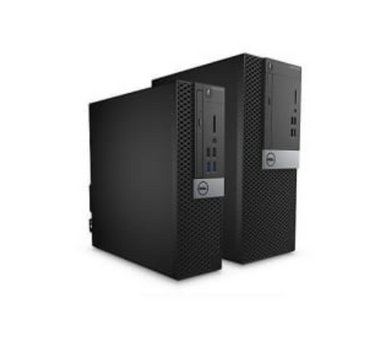
Performance PC
Meeting all the specifications needed, Dell desktop PC’s, accompanied by a large full HD monitor are available to be ordered along with the GelCount. These PC’s are equipped with GelCount software pre-installed and are configured to work flawlessly with the GelCount, therefore, providing the end user a full ‘turnkey’ solution.
Testimonials
We have now taken this best practice of industrialized colony counting and purchased the [GelCount] for our drug discovery activities at the Institute for Applied Cancer Science, University of Texas, MD Anderson Cancer Center.” “..the GelCount instrument has by far the most intuitive software and you don’t have to be a daily user to remember the workflow and settings etc.
Dr. Jannik N. Andersen,
UT MD Anderson Cancer Center, Houston, United States
We in Dr. Victor Levin’s Lab have found [the] GelCount [to be] as an absolutely fabulous option for counting clonogenic assays. The results are objective, reproducible and accurate. Earlier, we had been doing the same assays manually and now we are in a position to appreciate the amount of subjective error one could have. The most important output with this machine is the ‘volume’ [statistic] of the colonies in addition to the colony count. We truly recommend this magical piece of [equipment] to all those interested in doing clonogenic assays and other 3D studies.. It is cost effective and saves a whole lot of time.. go for it!
Dr. Sonali Panchabhai and Dr Yoshinori Kajiwara,
UT MD Anderson Cancer Center, Houston, United States
The detection and sorting algorithms seem very powerful and flexible, and the resolution at the high end is really quite remarkable. The ability to scale resolution down to what you need is a real time-saver, especially if you have large stained colonies. The high resolution capability is nice for tracking the growth progress of non-stained colonies as well.
Dr. Ryan Williams
UT MD Anderson Cancer Center, Houston, United States
The [Oxford] Optronix GelCount has been a fantastic addition to our laboratory setup, allowing rapid and efficient counting of clonogenic assays which would otherwise be a very time consuming and onerous process. The easy to use software reliably identifies colonies and greatly improves consistency in analysing these experiments. I would be happy to recommend the [Oxford] Optronix GelCount to other researchers.
Dr. Ross Carruthers
Institute of Cancer Sciences, University of Glasgow, Beatson Institute for Cancer Research, Glasgow, UK
I have used the GelCount for over 6 months. The machine reduces the analysis time considerably when compared to manual counting, and the results are similar between them. It has also the added bonus of reducing the subjectivity that could arise by manual counting, making the clonogenics more reliable. In addition, Oxford Optronix provides very good technical support. Overall, I would recommend this product as it reduces the time and increases the confidence in your results.
Dr. Natividad Gomez-Roman,
Institute of Cancer Sciences, University of Glasgow, Beatson Institute for Cancer Research, Glasgow, UK
With the GelCount from Oxford-Optronix I can finally forget about those many hours spent at the microscope trying to get a number of the amount of colonies in my soft agar experiments. Now I can easily image all the wells, and after choosing my desired settings, in a short time I can have the number of my colonies and their size. The GelCount allows me to follow the growing of the colonies in time by simply acquiring different images during the weeks, so that I can see the effects of the different sample treatments. Definitely, the use of the GelCount saves a lot of my time and now I can perform more experiments in a shorter time, with the additional advantage of acquiring a complete and clear image of the entire well for presentation purposes
Dr. Tiziana Scanu,
The Netherlands Cancer Institute (NKI-AVL), Amsterdam, The Netherlands
Specifications
| Physical | ||||||||||||||
|---|---|---|---|---|---|---|---|---|---|---|---|---|---|---|
|
Imaging method |
Visible light trans-illumination, high resolution 16-bit greyscale CCD line imager |
|||||||||||||
|
Imaging resolution |
user selectable between 300 – 2,400 ppi |
|||||||||||||
|
Depth of field |
Up to 5mm effective, above well base |
|||||||||||||
|
Sample loading method |
Removable loading tray, latching into a software-controlled motorized drawer |
|||||||||||||
|
Plasticware supported |
Multi-well plates (6, 12, 24, 48 and 96-well plates with/without lids; 35, 50/60 and 100mm Petri dishes with/without lids; selected T25 flasks |
|||||||||||||
|
Loading tray capacity |
With 6-, 12-, 24-, 48- and 96-well plates (up to 4 plates of any one type may be imaged simultaneously); with 35 mm (up to 24); 50 mm (up to 12); 100mm (up to 4); with certain T25 flasks (up to 8) |
|||||||||||||
|
PC interface |
USB 2.0 (x2) |
|||||||||||||
|
Dimensions |
560 mm x 450 mm x 155 mm (W x D x H) |
|||||||||||||
|
Weight |
20 kg (44 lbs) |
|||||||||||||
|
Power requirements |
100 – 240V ~ 1.5A, 50-60 Hz; 2 x T1.6A fuse |
|||||||||||||
|
Storage temperature |
10 – 40°C |
|||||||||||||
|
Operating temperature |
15 – 30°C |
|||||||||||||
|
Operating humidity |
0 – 70% (non condensing) |
|||||||||||||
| Performance | ||||||||||||||
|---|---|---|---|---|---|---|---|---|---|---|---|---|---|---|
|
Min. resolvable colony diameter |
Approximately 30 µm (at 2,400 ppi image resolution) |
|||||||||||||
|
Typical acquisition time |
12 minutes (four 6-well plates; 1,200 ppi) for non-adherent colonies |
|||||||||||||
|
Colony detection |
CHARM II™ (Compact Hough and Radial Map) image processing algorithm, dedicated to colony detection and size characterization, proprietary to Oxford Optronix Ltd. |
|||||||||||||
|
Supported colony types |
Adherent: stained (methyl blue, crystal violet, or equivalent). |
|||||||||||||
|
Variability in counting |
< 5% variability for repeated analysis of the same sample |
|||||||||||||
|
Numerical data output |
Automated exportation to Excel of colony counts, mean colony diameter, area, volume and other ‘statistics’ per well/dish. Optional ‘csv’ output of per-colony data and/or statistics histograms. |
|||||||||||||
|
Image output |
Per well/dish or compound bitmap images for general purpose importation/printing. Per well/dish raw images for off-line image analysis and archiving. |
|||||||||||||
Software Screen Shots
The GelCount features cutting-edge image analysis software presented within an intuitive and easy-to-use interface.
The subsequent screen views highlight some of the GelCount’s key software features:
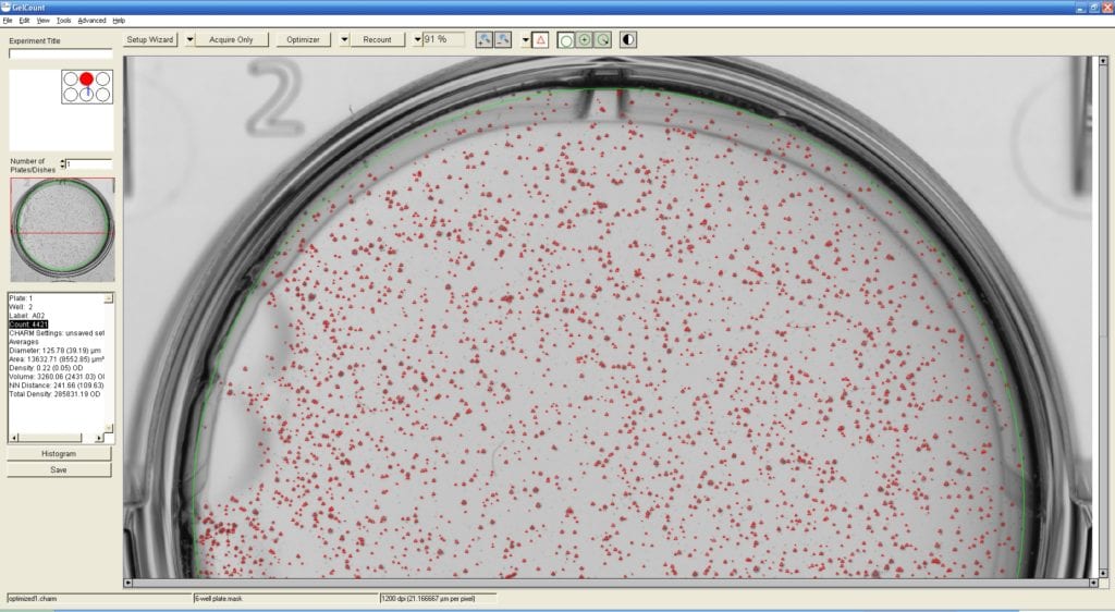
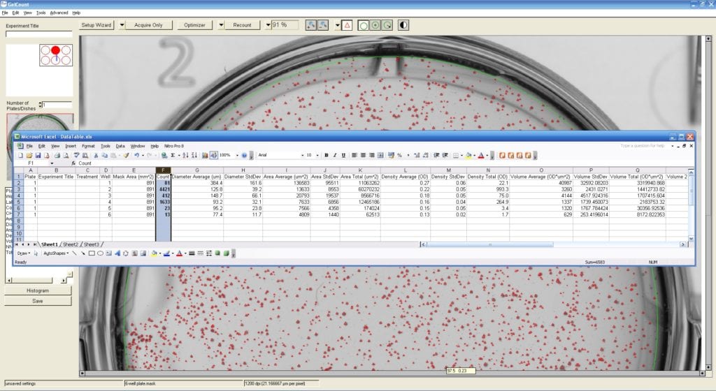
Multi-well plates are assessed one well at a time with an uncomplicated zoom and pan control providing easy image navigation. This allows any region of the well to be viewed quickly at high resolution for an up-close authentication of colony detection. All discovered colonies are highlighted by size-proportional red markers. The current colony count and other related colony statistics are presented in the summary panel in the left margin.
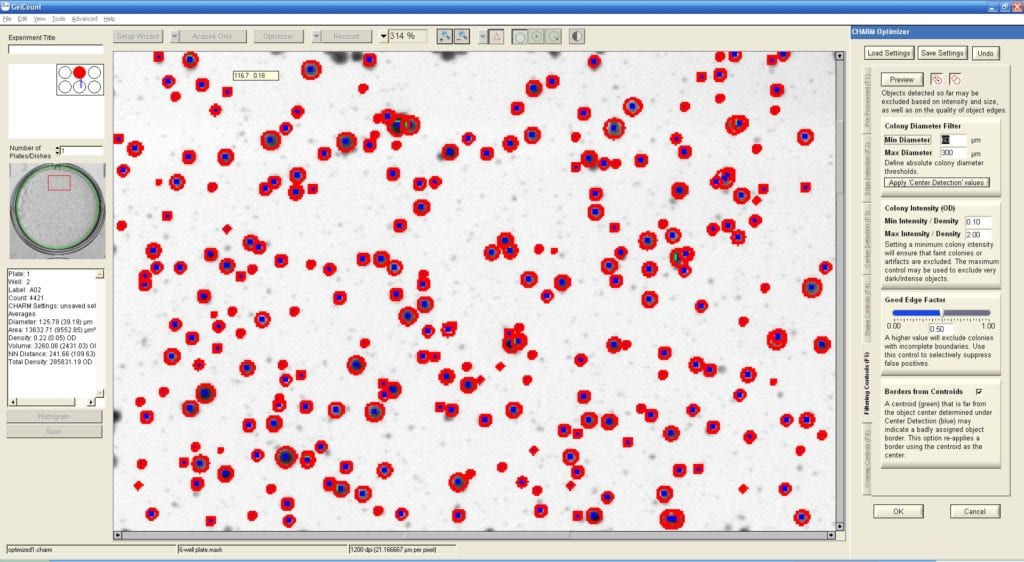
Colony detection is fully user-definable within the software from the “Optimizer” tab, which allows fine-tuning of several sensitivity and shape-related parameters and the capability to include or omit colonies from counting based on size. The effect of any modification in the tab is previewed instantaneously on the image and all parameters can be stored for re-use in future experiments. The GelCount achieves counting proportionality and reproducibility by objective and consistent application of the user-characterized settings.
All images of colony plates or wells can be saved as a general-purpose bitmap format (for publication or presentation quality images) or in a raw image format, which support the processing and re-processing of samples ‘off-line’ on any workstation with the installed GelCount software.
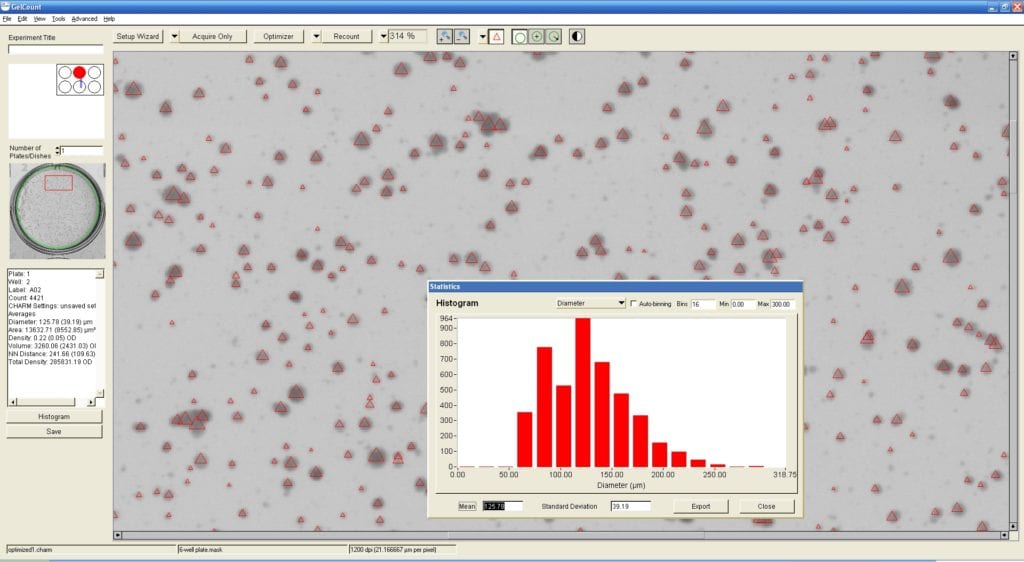
The GelCount is also efficient in calculating a unique set of statistical distributions, such as colony size, in a histogram form. Graphical data can be manually or automatically exported as a bitmap image or in Excel compatible raw data formats.
Colony detection is fully user-definable within the software from the “Optimizer” tab, which allows fine-tuning of several sensitivity and shape-related parameters and the capability to include or omit colonies from counting based on size. The effect of any modification in the tab is previewed instantaneously on the image and all parameters can be stored for re-use in future experiments. The GelCount achieves counting proportionality and reproducibility by objective and consistent application of the user-characterized settings.
Publications & Articles
Publication Highlight: Human Articular Chondrocytes Retain Their Phenotype in Sustained Hypoxia
Publication Highlight: Human Articular Chondrocytes Retain Their Phenotype in Sustained Hypoxia While Normoxia
Application Note: Physoxic Cell Culture – Yes or No?
Physoxic Cell Culture - Yes or No? Download PDF Here Oxygen is a
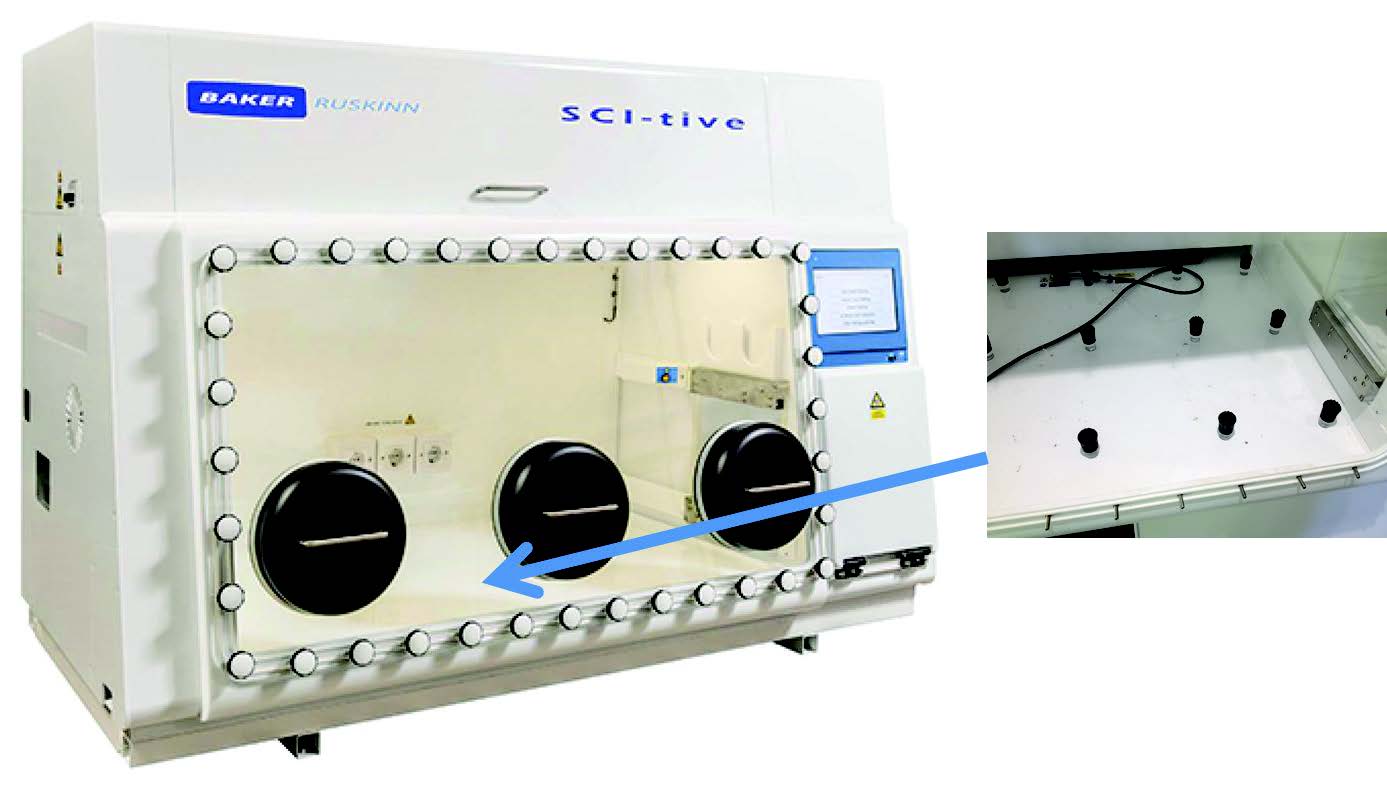
Increasing Cell Adherence with a Hypoxia Workstation
Increasing Cell Adherence with a Hypoxia Workstation System Used:SCI-tive Download PDF Here Abstract