Preclinical scientific imaging is an essential tool for studying disease mechanisms and investigating new therapies. It plays an important part in bridging the gap between the earliest phases of research and clinical trials, allowing researchers to visualize and study the anatomy and physiology of animal models of human disease.
Importance of Preclinical Scientific Imaging in Research
Medical research is an expansive field. It explores basic scientific principles that may expand non-clinical understanding and lead to pharmacological innovations that directly impact patient health and well-being. Preclinical scientific imaging tools apply to both. It is an essential component of pre- and non-clinical research helping researchers to study disease progression, therapeutic efficacy, potential toxicities, and more. Imaging data can also be used to develop biomarkers and guide the design of subsequent clinical trials.
Overall, preclinical scientific imaging covers a broad range of imaging modalities that provides valuable insights into disease mechanisms and helps to accelerate the development of new treatments.
Common Imaging Technologies Used in Preclinical Research
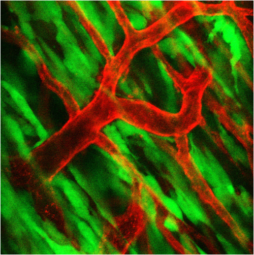
A wide variety of imaging technologies are used in preclinical research. Below are some of the most common imaging modalities used:
Magnetic Resonance Imaging (MRI)
MRI is the gold standard in soft tissue imaging. It is a non-invasive imaging technology that uses a powerful magnetic field and radio waves to produce high-resolution images of anatomical structures, physiological processes, and even molecular/cellular interactions.
Preclinical MRI applications vary widely, but it is routinely deployed to image anatomical and morphological targets, study neurological disorders, and monitor tumor progression.
Computed Tomography (CT)
CT imaging uses X-ray technology to recreate three-dimensional images of anatomical structures from a series of cross-sectional images. It is useful for visualizing bones, lungs, and other dense tissues, and can be used to monitor disease progression and assess the efficacy of therapies in preclinical models. Preclinical CT is often used in concert with other imaging techniques, such as MRI or positron emission tomography (PET).
Positron Emission Tomography (PET)
PET is a non-invasive imaging technology that uses a radioactive tracer to visualize metabolic activity in the body. PET is useful for visualizing tumors, tracking the distribution of drugs, and monitoring disease progression in preclinical models. Combined PET/CT systems often provide extremely high-performance imaging capabilities, enabling true depth of interaction for resolution uniformity and high sensitivity.
Bioluminescence Imaging (BLI)
BLI is a relatively novel imaging technology developed to non-invasively study ongoing biological processes. It uses light-emitting proteins to monitor gene expression, protein-protein interactions, and cellular processes in real-time. This makes BLI an extremely useful tool for studying infection, cancer progression, and gene expression.
Looking for Preclinical Scientific Imaging Solutions?
Preclinical research is a complex field. Scientists must sift through enormous amounts of information simply to choose the right equipment to fulfill their requirements. When setting up your study, how do you choose between the various preclinical imaging paradigms? Scintica was established with the simple goal of empowering researchers by simplifying solutions.
We are committed to providing our clients comprehensive support in various aspects of their research endeavours. This includes assisting with equipment usage, facilitating discussions on research objectives, and providing guidance on grant writing. We also engage in thought-provoking discussions on ongoing research projects, ensuring our clients can access the latest advancements in preclinical scientific imaging. Contact us today for more information on our preclinical imaging expertise.