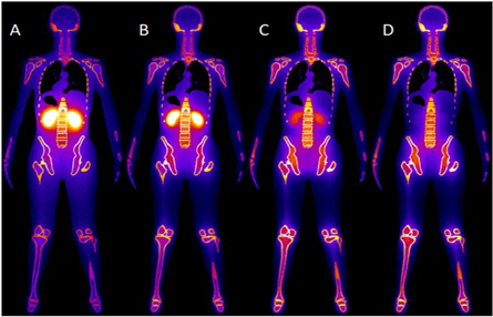
Certis Oncology Solutions is a precision oncology and translational science company. It works directly with cancer patients and their oncologists to help determine the best therapies for individuals, and also partners with pharmaceutical companies to help develop the next generation of anticancer therapies. Certis’s approach to studying drug efficacy is rooted in orthotopic patient derived xenograft (O-PDX) models. Because tumors are internal to the animal, they usually cannot be measured by calipers. Certis overcomes this challenge by using magnetic resonance imaging (MRI), employing the Aspect M3 Compact MRI to generate high-resolution 3D anatomical images to monitor anti-cancer therapies in real time.
Topics that will be discussed in this webinar include:
- Overview of precision oncology and testing platforms
- Comparison on subcutaneous and orthotopic in vivo models
- Imaging modalities for pre-clinical in vivo studies
- Applications of MRI in precision oncology and preclinical in vivo pharmacology studies
Interpatient tumor heterogeneity, wherein no two patients with the same subtype of cancer are genetically similar, has given rise to a clinical therapeutic approach known as precision oncology. Precision oncology is a practice where patients are given specific therapies tailored to their individual tumor, regardless of subclass. At its forefront is molecular profiling—a way to identify mutations and biomarkers that can often be used to narrow the field of potential therapies. But molecular profiling is a static characterization of the cancer, and alone cannot completely predict which drugs will—or will not—work for an individual patient. A functional assay, where a patient’s own living tumor is tested against an array of therapies, remains the most accurate method of determining which treatment would work best.
Science has long relied on mouse models to research human disease, and there is renewed interest in their application in precision medicine. Orthotopic Patient-Derived-Xenograft (O-PDX) models are the most clinically relevant models for cancer research available today.
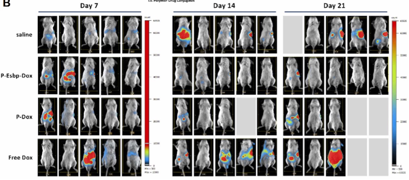
O-PDX models are created by engrafting human tumor tissue in the same anatomic location in immunocompromised mice (liver to liver, breast to breast, lung to lung, etc). Unlike commonly used subcutaneous models, O-PDX models retain human cancer’s complex physiology, making them more clinically relevant and highly predictive of therapeutic response, future metastases, and recurrence. Due to the internal implantation of tumors, advanced imaging modalities are required.