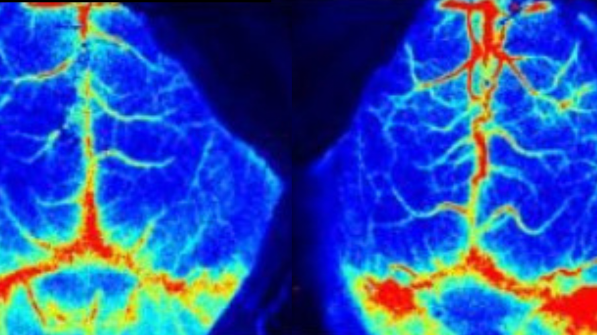
Using the Laser Speckle Imaging System to Observe Changes in Cerebral Blood Flow
System Used:
RFLSI-ZW Laser Speckle Contrast Imager

After Lymphatic Ablation and Quantitative Analysis
Background and Customer Needs
The circulatory system is a continuous closed system of ducts distributed throughout the body, including the cardiovascular system and the lymphatic system. What circulates in the cardiovascular system is blood. What flows through the lymphatic system is lymph. The lymphatic system can also be thought of as an auxiliary part of the venous system, as lymph flows centrally through a series of lymphatic canals that eventually drain into veins.
The brain doesn’t have its own lymphatic network, but the membrane around the brain, called the meninges, does have a network of lymphatic blood vessels. The meningolymphatic system was first discovered in 1787.
Extravasated erythrocytes in cerebrospinal fluid (CSF) critically contribute to the pathogenesis of subarachnoid hemorrhage (SAH). A subarachnoid hemorrhage means that there is bleeding in the space that surrounds the brain. It’s a very serious condition and can be fatal.
Meningeal lymphatics have been reported to drain macromolecules and immune cells from CSF into cervical lymph nodes (CLNs). However, whether meningeal lymphatics are involved in clearing extravasated erythrocytes in CSF after SAH remains unclear.
The Demand of the Research
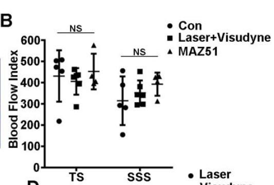
There are a set of results to tell these achievements, after the SAH model of C57BL/6, behavioral analysis, Visudyne treatment, flow cytometry, ICG-NIR imaging, tissue processing are all done to define the function of meningeal lymphatics after SAH, but the changes in cerebral blood flow after lymphatic ablation should be quantitatively analyzed to make the entire research completed, since there only three systems inside the brain, the lymphatic network, vascular system and the cerebrospinal fluid circulation.
How was The System Used
RFLSI Ⅲ provides a way to show the parts that do not produce or induce changes, thus removing interference with the results of the changes so that researchers can find the actual cause.
Results and Effects
With a series of experiments, a markedly higher number of erythrocytes are accumulated in the lymphatics of CLNs and meningeal lymphatics after SAH. When the meningeal lymphatics are depleted in a mouse model of SAH, the degree of erythrocyte aggregation in CLNs is significantly lower, while the associated neuroinflammation and the neurologic deficits are dramatically exacerbated.
In addition, during SAH lymph flow is increased but without significant lymphangiogenesis and lymphangiectasia.