Simultaneous Positron Emission Tomography (PET) and Magnetic Resonance Imaging (MRI) provides a unique non-invasive opportunity to image both the functional and structural characteristics of biological systems. PET imaging shows high-sensitivity to dynamic metabolic and functional processes through tracking of biomarkers in vivo but lacks the resolution to resolve detailed anatomy. MRI produces exceptional soft-tissue contrast but often lacks the ability to visualize metabolic processes. Combining both modalities allows clinicians and researchers to collect a wealth of biological information with regard to both  structure and metabolic processes in-vivo. Much recent technical development has focused on the integration of both modalities within a single system to allow for simultaneous collection, which leads to reduced scan time and near perfect spatiotemporal correlation between images. Of particular importance is developing these capabilities in the small animal pre-clinical setting providing invaluable opportunities to study biological processes and test therapeutic strategies.
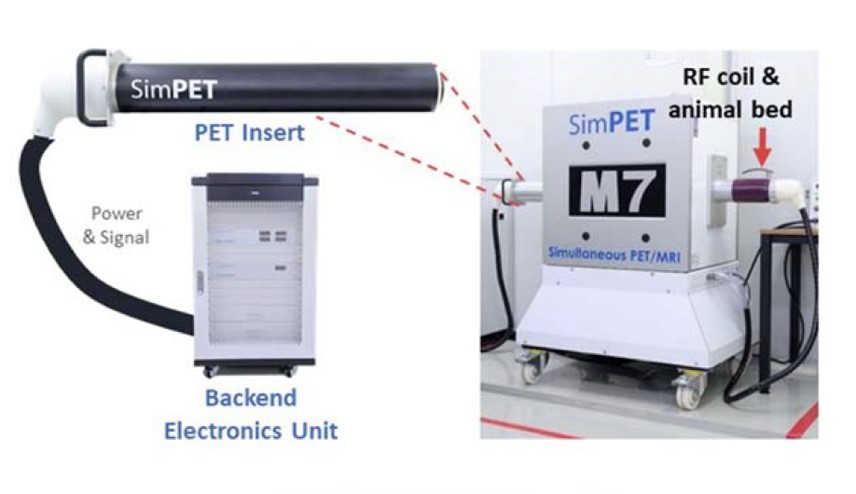
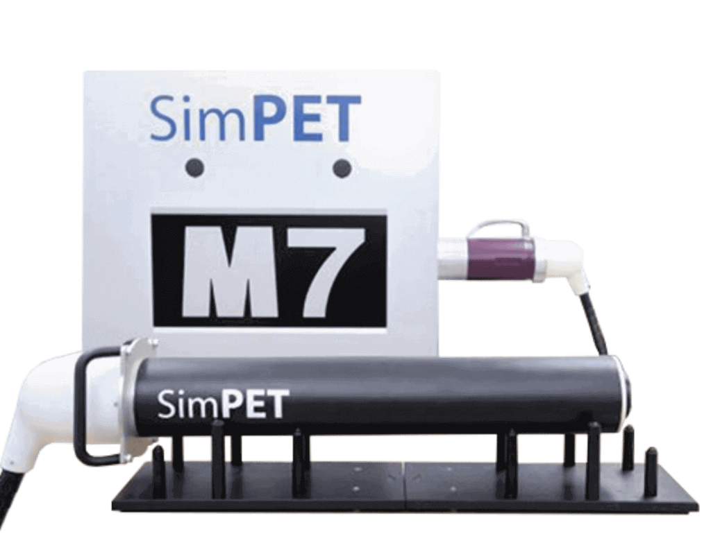
In this study, researchers at the Seoul National University College of Medicine present the performance evaluation of the SimPET/M7 system; an advanced simultaneous PET/MRI scanner dedicated to preclinical animal imaging. Following the NEMA NU 4-2008 standard (Performance Measurements of Small Animal Positron Emission Tomographs) the performance of the system was 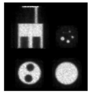 assessed. Further, compatibility of SimPET with simultaneous MRI was assessed using both an ex-vivo phantom and in-vivo mouse imaging. Here we present a brief summary of results.
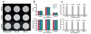
The spatial resolution at center based on a three-dimensional ordered-subset expectation-maximization (3D OSEM) reconstruction without and with warm background was 0.7 mm and 1.45 mm, respectively. Peak sensitivity was 4.21 % (energy window = 250–750 keV) and the peak noise equivalent count rate with the same energy window was 151 kcps at 38.4 MBq. As measured using the NEMA NU-4 image quality phantom, the uniformity was 4.42 %, and the spillover ratios in water- and air-filled chambers were 14.6 % and 12.7 %, respectively (shown to the right).
A hot rod phantom was used to assess the ability of the PET scanner to resolve spatially distinct objects. This phantom had six groups of rods with diameters of 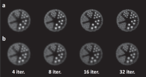 0.75, 1.0, 1.35, 1.7, 2.0 and 2.5 mm. The spacing from the center of each neighboring rod was twice it’s diameter. Shown are representative images obtained with a 3D OSEM reconstruction A) with the incorporation of resolution recovery using point spread functions and B) without point spread functions. In A the 0.75 mm rods are distinguishable and > 1 mm rods are clearly separated shown excellent image resolution.
As anticipated, different PET conditions and MRI pulse sequences did not appreciably affect the SNR and uniformity of MRI images and PET count rates representing excellent integration between the two subsystems (results shown below).
 To demonstrate the capabilities of the system in-vivo, simultaneous PET/MRI with 64Cu-NOTA-ironoxide was performed in a BALB/c mouse. In the images below we see excellent spatiotemporal correlation of the PET/MRI images 10-min post injection and strong signal intensity in the abdominal aorta highlighting the capabilities of simultaneous PET/MRI in-vivo.
While we have only presented a subset of the performance results here, they demonstrate the strong capabilities of the SimPET/M7 and highlight the potential for further research applications. The ability  to collect these two modalities simultaneously with limited interference provides an invaluable opportunity for researchers to gain a deeper understanding of a wide range of biological processes leading to improved diagnostic and prognostic capabilities.
For more information about the Aspect Preclinical MRI scanners with SimPET insert please contact Scintica Instrumentation.