Frequently Asked Questions : High-frequency preclinical ultrasound
This Frequently Asked Questions document has been designed to help answer some of the most commonly asked questions by people newer to high frequency ultrasound imaging, but also addresses some Prospect T1 specific questions. The questions have been divided into a few groups:
- Imaging application questions
- Ultrasound as an imaging modality
- Prospect T1 technical specifications
- Animal handling capabilities
- Contrast agent imaging
Imaging Application Questions:
1. What are the recommended applications for the Prospect T1?
- Cardiovascular Research
- Systolic and Diastolic function
- Blood flow velocity
- Pulsatility and Resistive Indices
- Vessel diameters
- Image guided injections
- Cancer Biology and Research
- Tumor detection
- Tumor volumes
- Tumor perfusion
- Therapeutic response
- Detection of metastasis
- Image guided injection for orthotopic tumor models
- Abdominal and Reproductive Organ Research
- Organ identification
- Organ size
- Pathological changes
- Organ perfusion
- Developmental Biology
- Confirmation of pregnancy
- Embryo staging
- Counting embryos
- Image guided injections into embryos
- Ophthalmic Research
- Anterior and posterior structures
- Retinal perfusion
2. What species can be imaged using the Prospect T1?
Traditionally the Prospect T1 has been designed for small animal imaging, for example on mice and rats, and similarly sized animals. There are 2 available animal handling tables, sized for mice and for rats. However, any animal of similar size can be positioned on these tables. The use of the Prospect T1 is however not limited to just these species and sizes, the main limitation of the system will be the penetration of the ultrasound waves to the target structure. For example, high-frequency images, with high resolution, can be acquired on larger species using the Prospect T1 probes as long as the structure is superficial – for example, carotid arteries in a rabbit, however of note is that these species will not fit on the animal handling platform. Additionally, other small animals such as zebrafish, amphibians (frogs, salamanders, etc.), and others, can be imaged using the Prospect T1.
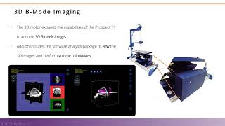
3. What applications is the 3D motor used for?
The 3D motor is used to acquire a stack of B-mode images across a defined range (maximum 30mm) with a given step size (minimum 0.05mm). These images are traditionally acquired on tumors, abdominal organs, muscles etc. The 3D motor is not triggered on the ECG signal, so is not appropriate for 3D imaging on the heart.
Ultrasound as an Imaging Modality
The most common applications for the Prospect T1 are listed below. However, almost all soft tissues, with the exception of the lungs (due to the air in the alveoli) and the brain (due to the skull) can be imaged:
4. Why is high-frequency ultrasound important for preclinical imaging?
When talking about ultrasound imaging, one of the most important considerations is the frequency of sound that is used to acquire the images.
As the frequency of sound increases, for example from 1 to 10 to 40MHz the resolution of the acquired images also increases; for example, a 40MHz probe on the Prospect T1 provides image resolution of 50µm. However, as frequency and resolution increase, the ability of the sound to penetrate the tissue decreases. This is why lower frequencies of sound are used to scan human subjects, while high frequency is favorable when scanning small animals such as mice and rats.
When we consider these smaller species, it is important to think about the size of the structures which will be imaged, and therefore the required resolution. However, one must also consider the depth of the imaging target as well. For example, the heart of a rat is quite deep compared to that of a mouse, and therefore one would select the 20MHz probe on the Prospect T1 system to image the rat heart, while a mouse heart could be imaged using the 40MHz probe.
Prospect T1 Technical Specifications
5. What scanning modes are available?
The standard software includes all available imaging modes, including:
- B-mode
- M-mode
- 4 Doppler modes: Pulsed-Wave Doppler mode, Color Doppler mode, Power Doppler mode, and Tissue Doppler mode
Additionally, there are some add-on hardware components available to expand the system’s capabilities, including
- 3D motor, allowing for 3D volume imaging
- Image guided injection mount
- Shear wave elastography
- Sonoporation probe, along with integration into the imaging software
6. What frequencies can each of the probes reach?
There are 3 different probes available on the Prospect T1 system, each probe has a center frequency, along with a range called the broadband frequency:
- PB506e: 30-60 MHz (50MHz center) 50MHz Probe, traditionally used on superficial structures2.
- PB406e: 30-60 MHz (40MHz center) 40MHz Probe, traditionally used on mice
- PB207e: 15- 30 MHz (20MHz center) 20MHz Probe, traditionally used on rats, and for non-linear contrast imaging
7. Can a keyboard and mouse be attached to the tablet to make data input and measurements on the system easier?
Yes, the system comes with both a keyboard and mouse to help make measurements, image labeling, and other data input easier. However, important to note that the 15” screen in a touch display which works well even with gloves on, providing the user several different ways to interact with the system.
8. Is offline analysis possible, and what are the data formats for export?
Yes. The software installed on the system includes the full complement of measurement capabilities. This software is also available for offline analysis on computers using the Windows operating system. This software is available as a per computer license.
Data can be exported in a .csv format for compilation and statistical analysis. The measurements can be completed multiple times on an image, the software will then calculate the average and standard deviation, as well as providing the individual measurements as well.
Images can be exported as both still and movie images for display to others. DICOM image exports are also available.
For images acquired in 3D, individual bmp files for each slice can be exported for analysis in third party software.
9. Can the digital radio frequency data be saved on the Prospect T1?
Yes. On the Prospect T1 the RAW digital RF data can be saved in all imaging modes, either by default or user selected when desired. This data can then be imported into third party software for further analysis. Scripts for MATLAB are available upon request.
Animal Handling Capabilities
10. Are physiological parameters measured on the Prospect T1?
Yes, the animal tables have built-in electrodes on the surface. The paws are attached to the electrodes allowing both the ECG and respiratory signals to be acquired and displayed with the images. Additionally, a rectal probe is used to monitor the temperature of the animals, while the table itself is heated to help maintain the core temperature of the animal while under anesthesia.
11. What anesthesia is typically used with the Prospect T1?
Anesthesia is suggested for ultrasound imaging to ensure the images are consistent and reproducible. The effects of anesthesia need to be considered when looking at functional measurements, such as cardiac function.
The Prospect T1 comes with an integrated inhaled anesthesia system, however the nose cone holder can be adapted to work with other anesthesia systems. Of course, injectable anesthesia can also be used with the system, if appropriate for the specific imaging study.
12. Is ECG and Respiratory gating possible?
Currently, the ECG signal can be used to acquire a retrospective B-mode images with a higher frame rate, used primarily for long and short axis images in B-mode. Corrections in 3D images caused by respiratory motion can be dealt with in volume measurements, during post-processing steps.
13. Can conscious animal imaging be performed with the Prospect T1?
Yes, it is possible to image conscious animals with the Prospect T1, for example to look at cardiac function. A confident animal handling person is necessary to ensure results are consistent and reproducible.
Contrast Agent Imaging
14. Do I need a contrast agent to acquire an image with the Prospect T1?
No, contrast agents are not needed to acquire images using the Prospect T1. The inherent properties of the tissue allow for the differentiation of various tissue types within the acquired images. For example, liquid, such as urine in the bladder, or bile in the gallbladder will appear black on an ultrasound image, while various soft tissues will have varied shades of grey and imaging of structures throughout the depth of the animal can be imaged. However, bone and air do not allow the sound to penetrate the
ough, causing a very bright signal from the first interaction with these structures, but nothing below can be visualized.
15. Can I use contrast agents with the Prospect T1?
Yes, contrast agents may be used to visualize different structures when using the Prospect T1. Contrast agents used with ultrasound are typically microbubbles. These agents, such as the USphere agents from Trust BioSonics sold by Scintica, have a phospholipid shell with a biocompatible gas core. These agents are very small, approximately 1.1-1.4µm in size, allowing them to mimic red blood cells. Agents may be used to study perfusion in various tissues, tumors, and structures; or the agents may be functionalized to allow different moieties to bind to their surface to target specific biomarkers accessible on the endothelial cell layer. When multi-modal imaging may be desirable there are various versions of the agents which have fluorescent dyes incorporated in the center or on the shell of the bubbles. Finally, there are agents with a positive charge on their shell to help load the microbubbles with DNA or RNA, and when combined with sonoporation these agents are used for targeted gene transfection at the site of sonoporation.
16. Are there contrast agent imaging modes on the Prospect T1?
- Linear Contrast Imaging – this can be completed using either the 40MHz or 20MHz probes. A baseline image is acquired before any contrast agent is injected. A new image is acquired as the contrast agent is injected. The baseline is subtracted from the images where the contrast agent was injected, the difference is colorized green indicating the presence of the contrast agent.
- Non-linear, or Harmonic Contrast imaging – this imaging is only possible when using the 20MHz probe. In this situation Harmonic imaging is activated, the probe will transmit at a lower frequency (13MHz) but will receive at a higher frequency (26MHz). Tissue signal is minimal when imaging the 1st harmonic signal, while when microbubbles are injected a very strong signal is seen from the 1st This imaging mode is very sensitive to the presence of the contrast agent.
In both modes, the analysis may be performed on multiple regions of interest, in which a time vs intensity curve is generated to study the perfusion kinetics when a non-targeted agent is used.
Targeted agents may also be used in either mode, to study the binding of the specific agent to the designated biomarker.