


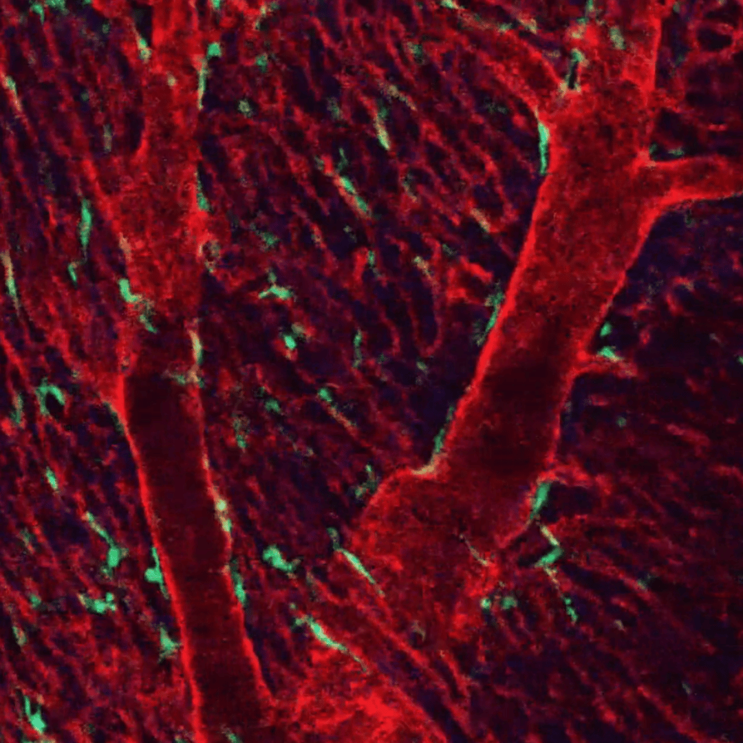
Article Highlight: Intravital Imaging of Cardiac Tissue Utilizing Tissue-Stabilized Heart Window Chamber in Live Animal Model
Article Highlight: Intravital Imaging of Cardiac Tissue Utilizing Tissue-Stabilized Heart Window Chamber in

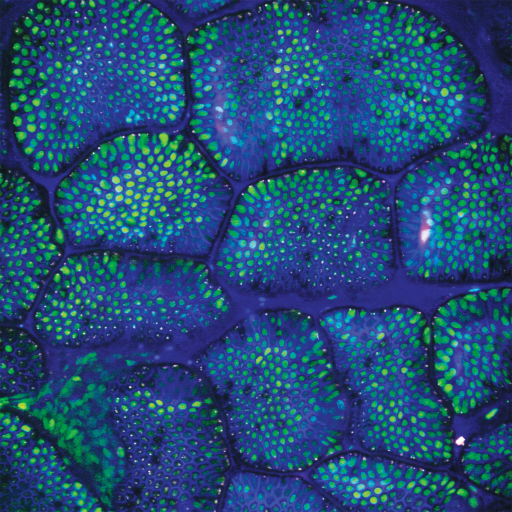
Intravital Microscopy- Real-time Tracking of Osteoclast Activity for Bone Disease Studies
Intravital Microscopy- Real-time Tracking of Osteoclast Activity for Bone Disease Studies View Paper
Application Note: Deep Brain Imaging Utilizing Novel Surgical Techniques: Cranial Imaging Window and Cannula with Intravital Tunable Two-Photon Microscopy
M-Series Application Note: Deep Brain Imaging Utilizing Novel Surgical Techniques: Cranial Imaging Window
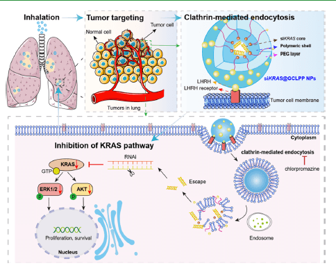
Publication Highlight : Inhalable siRNA Nanoparticles for Enhanced Tumor-Targeting Treatment of KRAS-Mutant Non-Small-Cell Lung Cancer
System Used: IVIM System M-Series Guolin Zhao, William Ho, Jinxian Chu, Xiaojian Xiong,
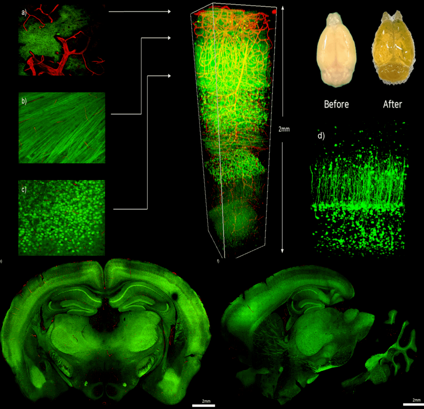
Leverage your research with IVIM Technology’s advanced imaging techniques for deep brain imaging
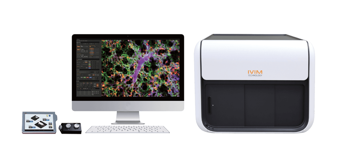
System Used In This Work: Intravital Microscopy (IVM) View Paper Here Introduction Our
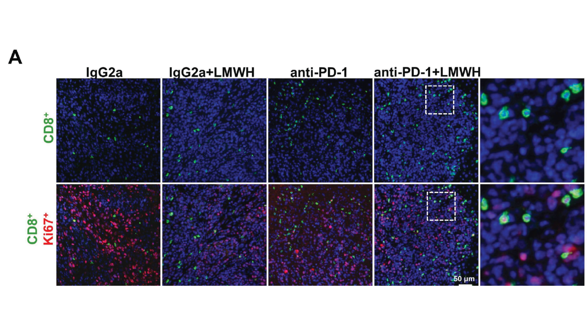
Low molecular weight heparin synergistically enhances the efficacy of adoptive and anti-PD- 1- based immunotherapy by increasing lymphocyte infiltration in colorectal cancer
Yibo Quan ,1 Jie He ,1,2 Qi Zou,1,2 Liuxi Zhang,1,2 Qihui Sun, 2