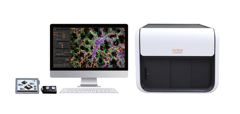

IVM is an all-in-one confocal/two-photon microscopy system designed and optimized for imaging live animal models.
The IVM line of products includes all-on-one two-photon, confocal, and dual-modal confocal and two-photon systems, specifically designed for longitudinal, in vivo, imaging of small animals. The enclosed and compact design of the system simplifies the installation and operation of the instrument as access to a dark room is not required. Additionally, the IVM systems are equipped with integrated animal physiological monitoring hardware to ensure the welfare of imaging subjects. Custom-made and free-space systems are available upon request for specific application such as retinal imaging.
Just talk to us about your model and your research needs!
– J. Ahn, et. al., Biomed. Optics Express, 10(6):2719 (2019)
– D.Y.Kim, et. al., Scientific Reports, 9:3560 (2019)
– J. Ahn, et. al., Biomed. Optics Express, 6(10):3963 (2015)
– K. Jung, et. al., Circulation Res., 112(6):891 (2013)
– J. K. Kim, et. al., Nature Protocols, 7(8): 1456 (2012)
– P. Kim, et. al., Nature Methods, 7(4):303 (2010)
– Z. Fan, et. al., Nature Medicine, 16(6):718 (2010)
Lung
– I. Park, et. al., European Respiratory Journal, 53:1800736 (2019)
– I. Park, et. al., Biomedical Optics Express, 9(5):2383-2393 (2018)
– S. Han, et. al., Science Translational Medicine, 8(335):335ra55 (2016)
Liver
– J. Moon, et. al., Biomed. Optics Express, 11(9):5132-5146 (2020)
– B. Oh, et. al., Diabetes, 67(3):473-485 (2018)
– Y. Hwang, et. al., Biomed. Optics Express, 8(10):4706-4716 (2017)
– A. L. van de Ven, et. al., Journal of Controlled Release, 158(1):148 (2012)
Small Intestine
– K. Choe, et. al., Journal of Clinical Investigation, 125(11) :4042 (2015)
– J. Ahn, et. al., Biomedical Optics Express, 6(10):3963-3972 (2015)
Colon
– J. W. Choi, P. Kim, et. al., Theranostics, 5(7):724-732 (2015)
– J. K. Kim, W. M. Lee, et. al., Nature Protocols, 7(8): 1456-1469 (2012)
– P. Kim, E. Chung, et. al., Nature Methods, 7(4):303-305 (2010)
Heart
– K. Jung, P. Kim, et. al., Circulation Research, 112(6):891-899 (2013)
Brain
– J. Lee, et. al., Biomed. Optics Express, 11(8):4835-4847 (2020)
Bone Marrow
– S. Ahn, et. al., PLos ONE, 12(11):e0187660 (2017)
Pancreas
– I. Park, et. al., Diabetes & Metabolism Journal, 44:193-198 (2020)
Spleen
– H. Choi, et. al., Science Advances, 6(15):eaa6980 (2020)
Prostate
– S. K. Ghosh, et. al., Cancer Research, 70(15):6119-6127 (2010)
Kidney
– E.M. Lee, et. al., Islets, 10(1):25-39 (2018)
– Z. Fan, et. al., Nature Medicine, 16(6):718-722 (2010)
Thyroid gland
– J. Y. Jang, et. al., EMBO Molecular Medicine, 9:750 (2017)
Mesentery
-K. Kim, et. al., Scientific Report, 6:33804 (2016)
Lymph Node
– K. Choe, et. al., Journal of Biomedical Optics, 18(3):036005 (2013)
– L. Zhi, P. Kim, et. al., Journal of Immunology, 187(5):2244-2251 (2011)
Eye / Retina
– J. Kim, et. al. Science Advances, 5(2):eaau6732 (2019)
– S. Ogura, et. al., JCI Insight, 2(3):e90905 (2017)
– J. Y. Lee, et. al., Invest. Ophthalmology & Visual Sci., 57(7):3390 (2016)
– J. R. Park, et. al., Invest. Ophthalmology & Visual Sci., 57:OCT331 (2016)
– D-Y. Park, J. Lee, et. al., Journal of Clinical Investigation, 124(9):3960 (2014)
Skin
– J. Ahn, et. al. Biomedical Optics Express, 9(8):3974-3982 (2018)
– J.Y. Kim, et. al., ACS Nano, 12(7):6904-6916 (2018)
– S. B. Kim, et. al., J. Cell Biology, 216(7):2201 (2017)
– Y. Hwang, J. Ahn, et. al., Optics Express, 22(10):11465-11475 (2014)
– J. Y. Lee, C. Park, et. al., Circulation, 122(14):1413-1425 (2010)
– X. Chen, P. Kim, et. al., PLoS ONE, 5, e13776. doi:10.1371 (2010)
Tumor Model
– J. Xu, et. al., Small, 14(50):1803601 (2018)
Circulating Tumor Cell
– J. W. Choi, et. al, Cancer Research, 75(21):4474-4482 (2015)
– H. Seo, et.al., Biomedical Optics Express, 6(6):2158-2167 (2015)
– D. Lee, et. al., Hepatology, 61(6):1978-1997 (2015)
Microcirculation / Blood Cell Flow
– I. Park, et. al., European Respiratory Journal, 53:1800736 (2019)
– I. Park, et. al., Biomedical Optics Express, 9(5):2383-2393 (2018)
– Y. Hwang, et. al., Biomed. Optics Express, 8(10):4706 (2017)
– J. Y. Jang, et. al., EMBO Molecular Medicine, 9:750 (2017)
– S. Han, et. al., Science Translational Medicine, 8(335):335ra55 (2016)
– K. Kim, et. al., Scientific Report, 6:33804 (2016)
– K. Choe, et. al., Journal of Clinical Investigation, 125(11) :4042 (2015)
– H. Seo, et.al., Biomed. Optics Express, 6(6):2158 (2015)
– J. W. Choi, et. al, Cancer Research, 75(21):4474 (2015)
– J. Ahn, et. al., Biomed. Optics Express, 6(10):3963 (2015)
– A. L. van de Ven, et. al., J. of Controlled Release, 158(1):148 (2012)
Drug Delivery / Efficacy Monitoring
– H. Choi, et. al., Science Advances, 6(15):eaa6980 (2020)
– I. Park, et. al., European Respiratory Journal, 53:1800736 (2019)
– J. Xu, et. al., Small, 14(50):1803601 (2018)
– J.Y. Kim, et. al., ACS Nano, 12(7):6904-6916 (2018)
– J. Ahn, et. al. Biomedical Optics Express, 9(8):3974-3982 (2018)
– K. Choe, et. al., Journal of Clinical Investigation, 125(11) :4042 (2015)
– A. L. van de Ven, et. al., J. of Controlled Release, 158(1):148 (2012)
Cell Delivery / Transplantation
– B. Oh, et. al., Diabetes, 67(3):473-485 (2018)
– E.M. Lee, et. al., Islets, 10(1):25-39 (2018)
– Z. Fan, et. al., Nature Medicine, 16(6):718-722 (2010)

