Science Blog

How Does In Vivo Optical Imaging Work?

Know more about this system

Vilber Webpage Newton 7.0 thumbnails 700x400
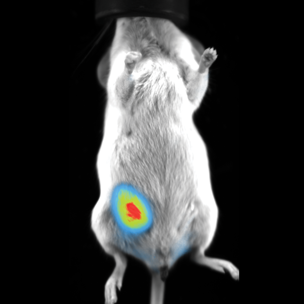
In vivo optical imaging has become crucial for scientists as they continue to research diseases and physiological processes through preclinical studies. This imaging method is commonly used in biomedical research because it is non-invasive and produces high-resolution images of biological tissues, organs, and processes in living animals at the molecular and cellular levels. In vivo imaging plays a key role in developing new drugs and treatments and evaluating their effects on their test subject. This blog post will look at the working principles of in vivo optical imaging and some of its preclinical applications. 

Working Principle

In vivo optical imaging utilizes the properties of light and how they react with biological tissues, as biological tissues can absorb, emit, or scatter light depending on their optical properties. A camera then captures this light, and the data is transferred to the instrument’s processing system to generate an image for visualization. It is important to note that in vivo imaging offers several techniques, including bioluminescence imaging, fluorescence imaging, and photoacoustic imaging. It can also use various types of light, such as bioluminescence, near-infrared, and visible light, and the most suitable techniques and light will depend on the imaging application.

Bioluminescence Imaging (BLI)

This imaging technique is used to detect luciferase-expressing or secreting molecules in the sample tissue and is commonly used in preclinical studies for cellular and molecular imaging in small animals because it is non-invasive and will not harm the test subject. Key benefits of BLI are that it offers a low signal-to-noise ratio (SNR), high sensitivity, and real-time visualization of biological processes.

Fluorescence Imaging

Fluorescence imaging is used when fluorescent reporter genes or dyes must be analyzed in vivo. It excites a target fluorescent molecule by utilizing an external light of a suitable wavelength, followed by a longer wavelength and lower-energy light to create an image of the sample. This technique is used extensively in preclinical studies, including protein interactions and protease activities.  

Photoacoustic Imaging

Photoacoustic imaging uses laser-generated light to irradiate sample material and then reconstruct the image of light to visualize biological tissues. It is currently used for studying cancer, cardiovascular diseases, microcirculation issues1, and more, as it can obtain functional, kinetic, molecular, and structural information about a sample. This method has proven to have vast potential in preclinical studies. 

Scintica and In Vivo Optical Imaging Solutions

Scientists have the challenging task of sifting through an extensive amount of information when choosing the most appropriate equipment for their research. Scintica was founded to provide researchers with the most suitable research tools and solutions and enable them to impact their field of work positively.   Scintica provides a range of instruments for in vivo bioluminescence and fluorescence imaging, which include state-of-the-art camera technology, integrated 3D tomography modules, and license-free acquisition and analysis software. Our Newton 7.0 includes several models for plant and small animal imaging studies and come equipped to meet various excitation and emission needs with optional accessories.   Contact a member of Scintica today to learn more about the vast possibilities in vivo optical imaging has on preclinical studies and more.

References

  1. https://www.ncbi.nlm.nih.gov/pmc/articles/PMC3262268/