
What does DFVS stand for?
DFVS stands for Doppler Flow Velocity System. It is a high-frequency, real-time pulsed Doppler measurement device designed specifically for measuring cardiovascular function in small animals.
What do you mean by Doppler?
The Doppler effect refers to the change in frequency of a wave in relation to an observer who is moving relative to the wave source so a Doppler ultrasound can estimate how fast blood flows by measuring the rate of change in its pitch (frequency).
How does it work?
Using small handheld probes, the DFVS operates quickly without the need for imaging to reach blood vessels with high accuracy. Flow velocities and differentials in various arteries, including the aorta, are reliably measured by placing the tip of the probe at a shallow angle relative to the direction of flow to be measured. High-frequency sound waves are bounced off the blood vessels and the returning sound waves (echoes) are picked up by the probe.
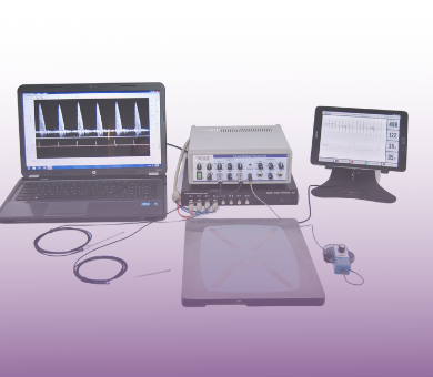
What components make up the DFVS system?
The DFVS is both compact and easily transportable, allowing it to be shared between multiple labs with ease. The four main components are:
- The handheld probe(s): emits and receives the sound waves to acquire a signal
- Transceiver: is used to change parameters of sound waves and receive the analog signals
- The Doppler signal digitizer: used for sampling and processing the Doppler signal
- The Doppler workstation: a laptop with custom software for analysis
Why not use cuff probes?
Cuff probes are invasive since the user is required to physically wrap the cuff around the vessel of interest. In contrast, the DFVS is completely noninvasive for small animals models allowing the user to acquire all desired signals longitudinally without surgical intervention. The DFVS works for larger animals, but requires the use use of invasive cuff probes.
What type of researcher could benefit from the DFVS system?
Anyone doing cardiovascular research could benefit from using the system. Specifically, groups interested in studying diastolic and systolic function, arterial stiffness, transverse aortic constriction (TAC), and coronary artery disease.
What type of measurements can you get from the DFVS?
- Cardiac systolic or diastolic function can be assessed under various conditions.
- Superior pulse wave velocity with simultaneous abdominal and aortic velocity measurements to assess arterial stiffness.
- Severity of transverse aortic constriction (TAC) can be assessed by comparing left and right carotid velocities.
- Pressure gradient across TAC can be estimated using stenotic jet velocity.
- Induced responses in microvascular beds can be measured through pulsatility and resistivity indices.
- Hyperemic velocity responses can be evaluated by assessing coronary flow reserve, a function of cardiac work affected by disease, age, and pressure/volume overload conditions.
- Banding effectiveness and coronary reserve measured with hyperemic/baseline velocity relations.
- Coronary flow distributions can be assessed through changes in the systolic/diastolic flow ratio.
What if I already have an ultrasound system?
The DFVS is still a valuable preclinical tool for those who already use ultrasound in their research. Specifically, pulse wave velocity and peripheral vascular measurements are more suited for a dedicated Doppler System due to the smaller probe size, allowing for smaller angles between the vessel of interest and probe.
For pulse wave velocity, the DFVS is also more beneficial because you can use more than one probe allowing the user to acquire simultaneous measurements of multiple locations. It is also typically faster to acquire signals from the DFVS compared to a traditional ultrasound system.
Can I acquire signals on animal models other than mice/rats?
YES, the DFVS has been used for a wide variety of animal models including guinea pigs, naked mole rats, zebrafish, giant danio, pigs, cats, and dogs. For larger animal models, measurements can be acquired using implanted silicon cuff transducers, with the leads tunneled subcutaneously.