iNSiGHT DXA

In Vivo Dual Energy X-Ray Absorptiometry (DXA/DEXA) For Preclinical Research.

"You won't regret purchasing this unit!"

Val Andrew Fajardo

Brock University

"Great instrument"

Jim McBride

Harvard School of Dental Medicine

System Overview

The iNSiGHT DXA system is a state-of-the-art in vivo Dual Energy X-Ray Absorptiometry (DXA/DEXA) technology designed for preclinical research. It provides a comprehensive solution for evaluating a wide range of metabolic disorders, such as osteoporosis, arthritis, diabetes, obesity, and musculoskeletal pathologies, including bone regeneration and muscle wasting diseases.

This system is equipped with a fully shielded X-ray cabinet, making it suitable for small animal DXA applications, with a focus on mice, rats, and animals of similar size (up to 500g). It provides fast and efficient body composition measurements. The system’s fast scan times of 25 seconds, combined with its low-dose radiation, make it ideal for longitudinal studies.

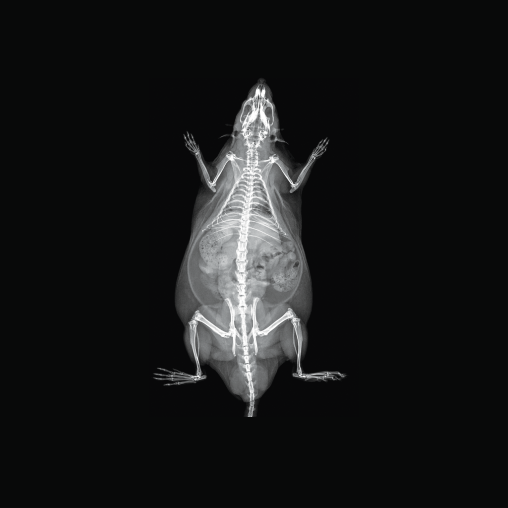
Representative rabbit DXA image generated with the iNSiGHT DXA system.
DXA image of a preclinical rodent showing skeletal structure and region of interest markers for bone mineral density and body composition analysis.

The iNSiGHT system uses dual energy x-rays to obtain highly accurate and reproducible body composition measurements. The system assigns each pixel to one of three compartments: fat mass, non-bone/lean mass, or bone mineral content, providing measurements such as bone mineral density, bone mineral content, bone area, tissue area, fat tissue as a percentage and weight, lean tissue as a percentage and weight, and total weight in grams.

This technology is non-invasive, non-destructive, and does not require any pre-treatment of the animal, contrast, or substrate injection. The iNSiGHT system also provides specific bone length measurements that can be drawn from the 2D x-ray image.

Features & Benefits

The iNSiGHT is a Dual Energy X-Ray Absorptiometry (DXA/DEXA) system designed specifically for in vivo preclinical research applications

Easy data acquisition

No preparation steps other anesthesia required. This can be done using the integrated anesthesia system, or with injectable anesthesia if desired.

Once ready the imaging subject is placed on stage and imaging can begin, using the easy-to-use acquisition software. 

Large scan area

The iNSiGHT system allows for a variety of animal models and tissue samples to be imaged, from 10g to 500g (scan area: 16.5 cm x 25.5 cm).

This allows users to image not only mice and rats, but other species such as non-human primates, fish, rabbits, etc.

25 second scan time

The iNSiGHT has the fastest in vivo scan time on the market, allowing for high-throughput imaging.

Comprehensive set of images and parameters

In a single scan (25 seconds) the x-ray attenuated image, bone mineral density map and color map are generated. 

A number of parameters are calculated from these images, either from the whole animal or for user defined regions of interest (ROI):

  • Bone Mineral Content (BMC) (g)
  • Bone Mineral Density (BMD) (g/cm2)
  • Bone area (cm2)
  • Tissue area (cm2)
  • Fat tissue (mass and percentage) (g & %)
  • Lean tissue (mass and percentage) (g & %)
  • Total mass (g)

Low-dose radiation

The iNSiGHT was designed to minimize the ionizing radiation (0.66 mGy) required to generate images and data. This minimization prevents any effect of the image acquisition on the subject, allowing for multiple imaging time points over the course of a longitudinal study.

Adjustable field of view and resolution

The iNSiGHT system has several levels of magnification, adjust the field of view (FOV) and resolution to suite your specific needs.
(pixel resolution from 100µm up to 31µm in Digital Radiography Mode)

Models & Specifications

iNSiGHT DXA preclinical imaging system.
Add-ons

iNSiGHT

DXA
Technical Specifications
  • Scan Method: Cone Beam
  • Animal Size: Mice, rats, and animals of similar size (up to 500g)
  • Image Area (maximum): 16.5 x 25.5cm
  • Pixel Size: 100µm at 1.2X 31µm at 4X with DR Mode
  • Operating System: Windows 10 and 11, 64bit
  • Dimensions (WxDxH): 66 x 61 x 113 cm
  • Power: 110/240VAC, 50/60Hz, 200VA
  • Operating Temp: 10 ~ 40oC

Applications

Past Webinars - iNSiGHT

Publications & Articles

Preclinical Bone & Body Composition Imaging

iNSiGHT DXA

Preclinical in vivo DXA/DEXA for bone mineral density, bone mineral content 

Measure BMD, BMC, fat mass, lean mass, and related body-composition endpoints from a 25-second scan. Low-dose X-ray acquisition supports repeated longitudinal measurements in small-animal studies.

iNSiGHT DXA preclinical imaging system.

iNSiGHT DXA Resources

Access product documents, technical information, FAQs, and educational resources for the iNSiGHT DXA system.
Product Documents
Brochure, validation white paper, and reference list.
FAQ
Review common questions about iNSiGHT DXA use and capabilities.
Specifications
Review system specifications, scan area, resolution, and configuration details.
Webinars
Watch iNSiGHT DXA presentations, software demonstrations, and research webinars.

iNSiGHT DXA System Overview

Representative rat DXA image generated with the iNSiGHT DXA system.
Representative rabbit DXA image generated with the iNSiGHT DXA system.

The iNSiGHT DXA system is a state-of-the-art in vivo Dual Energy X-Ray Absorptiometry (DXA/DEXA) technology designed for preclinical research. It provides a comprehensive solution for evaluating a wide range of metabolic disorders, such as osteoporosis, arthritis, diabetes, obesity, and musculoskeletal pathologies, including bone regeneration and muscle wasting diseases.


This system is equipped with a fully shielded X-ray cabinet, making it suitable for small animal DXA applications, with a focus on mice, rats, and animals of similar size (up to 500g). It provides fast and efficient body composition measurements. The system’s fast scan times of 25 seconds, combined with its low-dose radiation, make it ideal for longitudinal studies.

Key Imaging & Workflow Benefits

From fast, low-dose acquisition to quantitative bone and body-composition analysis, iNSiGHT DXA is designed to support efficient longitudinal preclinical imaging workflows. 

Fast Acquisition
25 Seconds
Scan Time
Quantitative Outputs
BMD + BMC
Bone, fat, lean & total mass
Longitudinal Study Fit
0.66 mGy
Low-dose, fully shielded acquisition
Regional Analysis
Whole + ROI
Whole-animal or user-defined regions
Simple Acquisition
No Contrast
No pre-treatment or substrate injection required

DXA Acquisition & Quantitative Analysis

iNSiGHT uses dual-energy X-rays to classify tissue into fat mass, non-bone/lean mass, and bone mineral content. From a single 25-second scan, the system generates quantitative bone and body-composition measurements for the whole animal or user-defined regions of interest. 

Bone Measurements

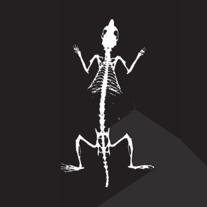
Bone mineral density, bone mineral content, bone area, and specific bone-length measurements drawn from the 2D X-ray image.

Rat bone mineral density image generated with the iNSiGHT DXA system.

Body Composition

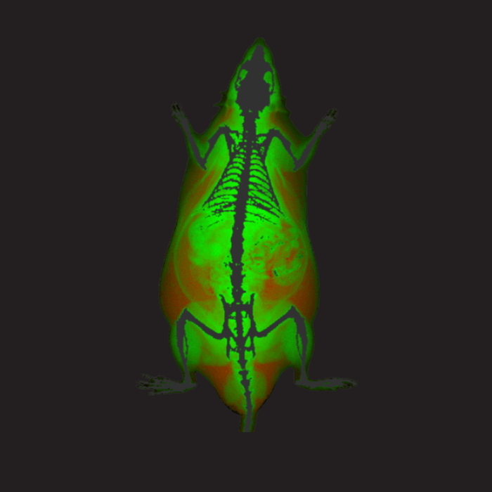
 Quantifies fat tissue mass and percentage, lean/non-bone tissue mass and percentage, tissue area, and total mass.

Rat body composition color image generated with the iNSiGHT DXA system.

Whole-Animal & ROI Analysis

Measurements can be calculated from the whole animal or from user-defined regions of interest.

DXA image of a preclinical rodent showing skeletal structure and region of interest markers for bone mineral density and body composition analysis.

DXA Image Outputs

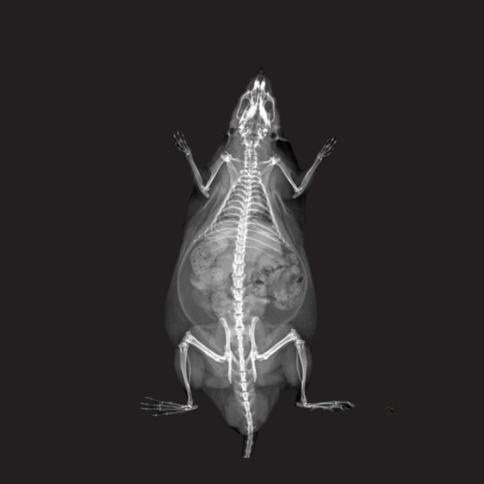
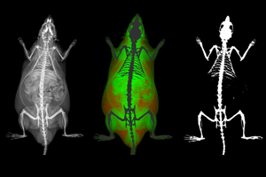
A single scan generates the X-ray attenuation image, bone mineral density map, and color map. 

Rat X-ray attenuation image generated with the iNSiGHT DXA system.

Applications in Preclinical Research

iNSiGHT DXA supports longitudinal assessment of bone and body composition across a range of preclinical disease models and study designs.

Metabolic Disorders

Metabolic disorders such as diabetes, metabolic syndrome, and obesity can alter body composition, energy metabolism, and musculoskeletal health. Small-animal models are commonly used to investigate the biological pathways involved in these conditions.

iNSiGHT DXA enables longitudinal assessment of body composition by measuring changes in fat mass, lean mass, bone mineral content, and related endpoints over time. This allows researchers to track disease progression or response within the same study. 

Measurements Relevant to Metabolic Research

  • Fat mass and percentage
  • Lean/non-bone mass and percentage
  • Bone mineral content
  • Total mass
  • Longitudinal changes over time
iNSiGHT DXA image used for the metabolic disorders application example.
Study Example

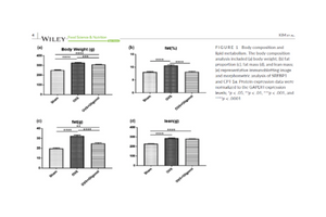
Diabetic mouse model

In the cited study, diabetic mice showed lower fat mass, lean mass, and bone mineral content compared with healthy controls.

Kishi et al., Exp Anim. 2023 · View study

Drug Safety & Toxicology

Drug safety and toxicology studies are an important step before target compounds advance to later preclinical studies or clinical trials. In addition to efficacy, researchers may assess potential toxic effects through changes in bone, fat, and lean tissues.

iNSiGHT DXA can assess body-composition changes following administration of a target compound at an effective dose, including changes in bone mineral density, bone mineral content, fat mass, and lean mass over time.

Measurements Relevant to Drug Safety & Toxicology

  • Bone mineral density
  • Bone mineral content
  • Fat mass
  • Lean mass
  • Longitudinal changes over time
iNSiGHT DXA image used for the drug safety and toxicology application example.
Study Example

Ovariectomized rat model

The cited study examined the effect of oligonol, a lychee-derived polyphenol, on skeletal muscle and body composition in ovariectomized rats.

Kim et al., Food Science & Nutrition. 2022

Musculoskeletal Diseases

Musculoskeletal diseases include pathologies affecting bone or muscle, including bone fractures and healing, bone and/or muscle regeneration, and muscle wasting diseases. Small-animal models can use DXA imaging to investigate genetic, molecular, and biochemical pathways involved in these conditions.

iNSiGHT DXA can assess disease progression or regression in response to a therapeutic regimen by measuring changes in bone mineral density and bone mineral content, as well as changes in fat and lean mass over time.

Measurements Relevant to Musculoskeletal Research

  • Bone mineral density
  • Bone mineral content
  • Fat mass
  • Lean mass
  • Longitudinal changes over time
iNSiGHT DXA image used for the musculoskeletal diseases application example.
Study Example

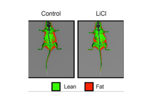
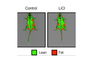
Low-dose lithium supplementation

In the cited study, low-dose lithium supplementation promoted adipose tissue browning and sarco(endo)plasmic reticulum Ca2+ ATPase uncoupling in muscle.

Geromella et al., J Biol Chem. 2022 · View study

Metabolic Bone Disease & Arthritis

Metabolic bone diseases include conditions that cause abnormalities or deformities in bone structure, including osteoporosis, rickets, osteomalacia, and osteogenesis imperfecta. Arthritis refers to joint disease and inflammation, with osteoarthritis and rheumatoid arthritis among the most common forms.

iNSiGHT DXA can assess disease progression or regression in response to therapy by measuring changes in bone mineral density and bone mineral content in affected joints. The source notes that these changes may be detected earlier in disease progression than on traditional X-ray images.

Measurements Relevant to Bone & Joint Research

  • Bone mineral density
  • Bone mineral content
  • Changes in affected joints
  • Whole-body BMC and BMD
  • Lumbar vertebral BMC and BMD
iNSiGHT DXA image used for the metabolic bone disease and arthritis application example.
Study Example

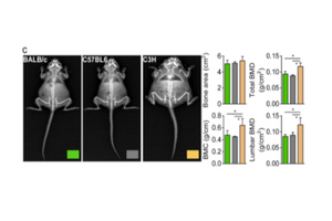
C3H mouse model

In the cited example, iNSiGHT DXA revealed higher BMC and BMD values in the whole body and lumbar vertebrae of C3H mice.

Kim MY et al., Exp Mol Med. 2021 · View study

Hypoxia

Hypoxia exposure in vivo is thought to alter the metabolic state of the exposed animal and may affect glucose metabolism and energy expenditure. Small-animal models are used to study these responses, including changes in body composition.

iNSiGHT DXA provides body-composition analysis during hypoxic exposure, with colorimetric imaging used to differentiate fat from lean tissue. This application is relevant to studies in altitude medicine, metabolic health, and cardiovascular research.

Measurements Relevant to Hypoxia Research

  • Fat tissue
  • Lean tissue
  • Body-composition changes during hypoxic exposure
  • Colorimetric fat/lean differentiation
iNSiGHT DXA image used for the hypoxia application example.
Study Example

Hypoxic exposure in mice

In the cited example, hypoxic exposure was associated with reduced fat mass in mice and increased energy expenditure through greater carbohydrate oxidation.

Park et al., BioMed Research International. 2020

iNSiGHT DXA preclinical imaging system.

Preclinical Imaging Workflow & System Features

iNSiGHT DXA combines streamlined animal handling, fast acquisition, and flexible imaging settings to support longitudinal preclinical studies.

Easy Data
Acquisition

No preparation steps other than anesthesia are required. Researchers can use the integrated anesthesia system or injectable anesthesia, if desired. Once prepared, the imaging subject is positioned on the stage and acquisition can begin using the acquisition software.

Flexible Animal & Sample Range

iNSiGHT supports animal models and tissue samples from 10 g to 500 g, with a maximum scan area of 16.5 × 25.5 cm.

25-Second
Scan Time

A 25-second scan time supports efficient, high-throughput imaging workflows.

Low-Dose Longitudinal Imaging

iNSiGHT is designed to minimize ionizing radiation, with a stated dose of 0.66 mGy, supporting multiple imaging time points over the course of a longitudinal study.

Adjustable Field of View & Resolution

Multiple magnification levels allow researchers to adjust the field of view and resolution for specific imaging requirements, with pixel resolution from 100 µm down to 31 µm in Digital Radiography Mode.

Shielded, Streamlined Workflow

The system uses a fully shielded X-ray cabinet and does not require pre-treatment, contrast, or substrate injection for imaging.

Configuration & Add-ons 

Configure iNSiGHT for Your Workflow

iNSiGHT DXA is available with optional add-ons to support anesthesia, higher-resolution imaging, and offline analysis.

Anesthesia Control

Anesthesia Control is available as an add-on for imaging workflows. Scintica also offers anesthesia systems for small-animal research.

Digital Radiography Mode

Collect higher-quality images with a pixel resolution of 31 µm.

Offline Analysis Software

Available as an add-on for iNSiGHT DXA. For broader biomedical image analysis, Scintica also offers Imalytics Preclinical.

Need help building the right imaging workflow?

Scintica offers complementary small-animal anesthesia and biomedical image-analysis solutions alongside iNSiGHT DXA. Explore the related systems below or contact our team to discuss your study requirements.

Technical Specifications

Key imaging, animal-size, and system requirements for the iNSiGHT DXA platform

Imaging & Acquisition
Main Use Case
Preclinical in vivo DXA/DEXA for bone and body composition measurements
Modality / Capabilities
DXA / DEXA; bone mineral and body composition analysis
Scan Method
Cone Beam
Pixel Size
100 µm at 1.2X; 31 µm at 4X with Digital Radiography Mode
Animal & Scan Area
Animal Size
Mice, rats, and animals of similar size (up to 500 g)
Maximum Image Area
16.5 × 25.5 cm
System Requirements
Operating System
Windows 10 and 11, 64-bit
Dimensions (W × D × H)
66 × 61 × 113 cm
Power
110/240 VAC, 50/60 Hz, 200 VA
Operating Temperature
10–40 °C

iNSiGHT DXA Imaging Gallery

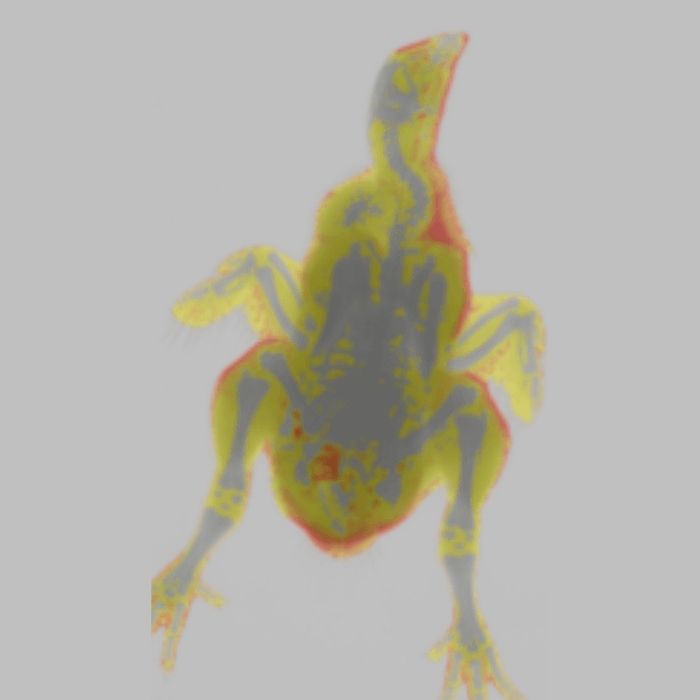
Explore representative iNSiGHT DXA outputs from preclinical models, including X-ray attenuation images, body-composition color images, and bone mineral density maps across rat, marmoset, and chicken studies. 

RAT

X-Ray Attenuation Image

MARMOSETS

DXA Imaging of Marmosets

RAT

Color Image

RAT

Bone Mineral Density Image

CHICKEN

Color Image

Resources

Continue Exploring iNSiGHT

Access product documentation and explore the latest webinars, publications, application notes, and articles related to the Rodent Surgical Monitor

Product Documents
Download brochures, specifications, validation documents, and other resources for this product.

Get the
DEXA
Brochure Immediately!

You will get IMMEDIATE access to the brochure via email, after completing the form.

Name