Having the ability to monitor cell culture over an extended period of time, offers insight into cell dynamics and function. Live cell imaging microscopes open up exciting and unique avenues, to analyze cell viability, health, migration, and even responses to external factors.
To support investigators in the life sciences to continue developing the understanding of cellular processes, The CytoSMART Omni has been developed as an automated bright-field lab microscope that visualizes whole culture vessels and can even be used within standard CO2-incubator. With the Omni researchers can perform kinetic assays by creating time-lapse videos that depict cellular behavior for days or weeks at a time.
BROCHURE
WHITE PAPER
REQUEST INFO
FREQUENTLY ASKED
QUESTIONS
BROCHURE
Omni FL
Omni FL is a live-cell imager capable of producing high-quality, whole-well brightfield or high-throughput fluorescence images of living cells. Equipped with a brightfield and two fluorescence channels (green and red), the CytoSMART® Omni FL can be used for continuous live-cell imaging, as well as endpoint assays. The applications of the CytoSMART® Omni FL range from a high-throughput analysis of cell viability and colony formation to the evaluation of transfection efficiency and co-cultures. The versatility and flexibility of the device are further enhanced through its ability to scan multiple types of transparent cell culture vessels, ranging from T75 flasks and 96-well plates to microfluidic chips and custom culture vessels. The intuitive and open design of the CytoSMART® Omni FL ensures that the device is easy to use and maintain, as well as it allows the incorporation of the device into established automated workflows.
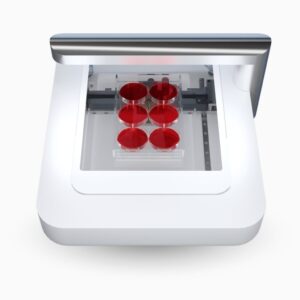
Omni
Being able to monitor cell cultures over time provides a great insight into their physiology and function. Live-cell imaging microscopes open up novel and exciting avenues to study cellular health, viability, colony formation, migration, and cellular responses to external stimuli.
To help life science researchers improve their understanding of cellular processes, CytoSMART Technologies has developed an automated brightfield microscope that can visualize an entire surface of a cell culture vessel and operate from inside a standard CO2-incubator, biological safety cabinet, or on a benchtop. Not limited to a specific type or quantity of culture vessels, the CytoSMART Omni captures cellular behavior by creating high-quality time-lapse videos for days or even weeks at a time.
 Real-time insight into cellular processes
Time-lapse imaging allows for researchers to identify important events in the progression of the cell culture across experiments. Researchers are now able disseminate events like attachment and detachment rates, cell death and even compare growth rates. Data collection using video monitoring allows users to capture changes within samples and compare the rate of change between samples. Further the integrated software makes seeing and graphing these changes a breeze as well.
Perform analysis at desired culturing environment
The CytoSMART Omni is designed for an incubator environment. Live-cell imaging systems require environmental control during the experiment. To ensure the proper environmental conditions, users can place the compact Omni inside any standard cell culture incubator. The smooth curvatures of the Omni platform ensure minimal airflow disturbance. This makes certain that your samples stay in their desired, homogeneous environment throughout the entire experiment.
Sample stability while imaging
Obtaining images is often highly disturbing to living cells. Environmental shock, like sudden changes in the atmosphere and mechanical disturbance of moving media can negatively influence cell viability. The Omni rapidly captures bright field images in cell-level detail without disturbing the cells. Researchers can set the desired imaging parameters using their own computer and the Omni will follow that scanning regime. At no point after placing the cells on the sample stage does the culture need to be moved or taken out of the incubator.. Imaging using such a stable system prevents unwanted disturbances and even has been shown to reduce imaging artifacts. What this means to the end user is automated time-lapse imaging eliminates the need for any manual interference and ensures reproducibility.
Complete overview of sample heterogeneity
Manual handling and cell seeding can cause a variable density distribution within culture vessels. Randomly selecting several areas of interest or tile scanning is common practice to overcome that issue, however this task time-consuming to set-up or post-process. The Omni automatically scans complete well surface areas and instantly stitches these images to give users a complete overview of cell coverage. It is the simplest device on the market to see differences in heterogeneity.
High throughput assays or cell line production
Cell based assays and subculturing will always benefit from standardization. Choosing the right vessel for your experiment and going through the process of identifying how cells behave in the vessel is critical for reproducibility. The Omni is flexible and is compatible to any transparent vessel that is lower than 55 mm. Any clear vessel with a surface area smaller than 99*131 mm can be imaged in full with the Omni.
Whole well time-lapses at cell level detail.
The CytoSMART Omni creates time-lapse videos in cell level detail; Whole well images are produced every hour by scanning the natural, unstained, culture vessel surface and automatically stitching the images.
The system is used to image various kinetic assays including cell proliferation, migration, and colony formation. Image analysis can be used to extract valuable information from time-lapse videos. Use the cell monitoring software to examine and compare parameters like cell confluence, area infiltration (wound healing assays) and growth rate across wells on a single plate.
[/fusion_tab][fusion_tab title=”Models & Specifications” icon=””][fusion_table fusion_table_type=”1″ fusion_table_rows=”” fusion_table_columns=”” hide_on_mobile=”small-visibility,medium-visibility,large-visibility” class=”” id=”” animation_type=”” animation_direction=”left” animation_speed=”0.3″ animation_offset=””]
| Model | Omni FL | Omni |
|---|---|---|
| Algorithms | Fluorescence: cell confluence and fluorescent object count & Brightfield: cell confluence, scratch assay, and colony assay | Brightfield: cell confluence, scratch assay, and colony assay |
| Unit Dimensions | 392 x 345 x 171 mm | 392 x 345 x 171 mm |
| Weight | 9 kg | 9 kg |
| Optics | Green and red fluorescence channels & Bight field with digital phase contrast | Bight field with digital phase contrast |
| Magnification | 10x fixed objective | 10x fixed objective |
| Light Source | LED | LED |
| Camera | 6.4 MP CMOS | 6.4 MP CMOS |
| Scan Area | 86×124 mm | 86×124 mm |
| Exported Formats | JPG, TIFF, XLSX, MP4 | JPG, XLSX & MP4 |
| Culture Vessels | Well-plates, petri dishes, flasks, microfluidic chips, and custom culture vessels (lower than 55 mm (2.2’’)) | Petri dishes, T25 – T225, triple flasks, and HYPERFlasks (lower than 55 mm (2.2’’)) |
| Operating Environment | 5 – 40 °C, 20 – 95% humidity | 5 – 40 °C, 20 – 95% humidity |
| Well Plates | 6 – 384 well plates | 6 – 384 well plates |
[/fusion_table][/fusion_tab][/fusion_tabs][/fusion_builder_column][/fusion_builder_row][/fusion_builder_container]