

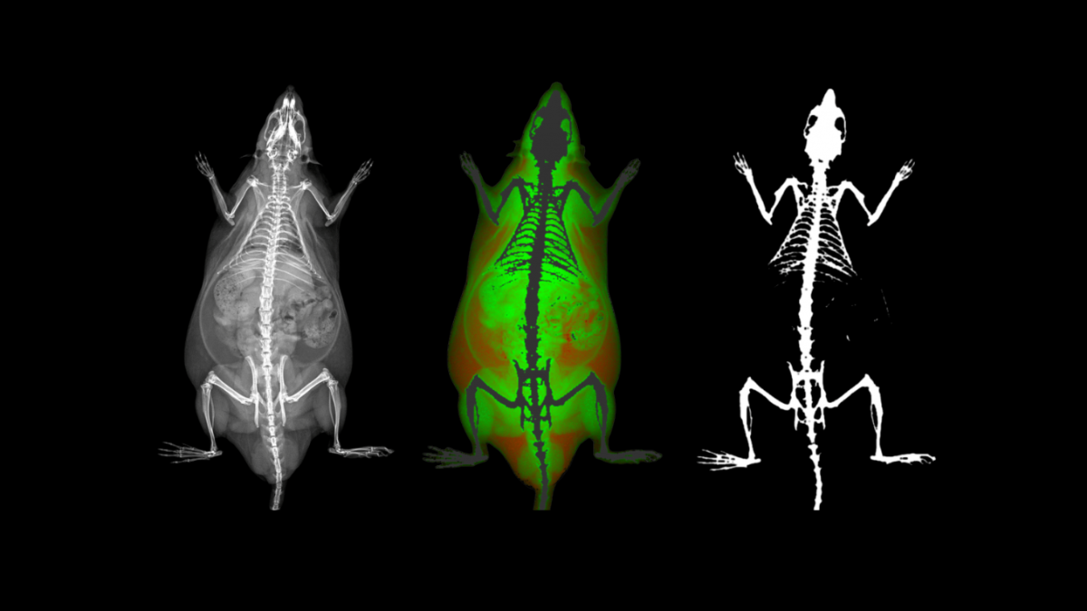
The Role of X-ray Absorptiometry in Preclinical Research
Preclinical research is being revolutionized by advanced imaging technologies. As a critical phase

University of Greenwich – Biological Imaging at Medway with Scintica
Add Your Heading Text Here Symposium on Biological Imaging at Medway with Scintica
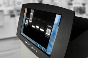
Gel Documentation Systems: Bridging the Gap in Modern Lab Research
A gel documentation system (GDS) captures images of nucleic acids and proteins in