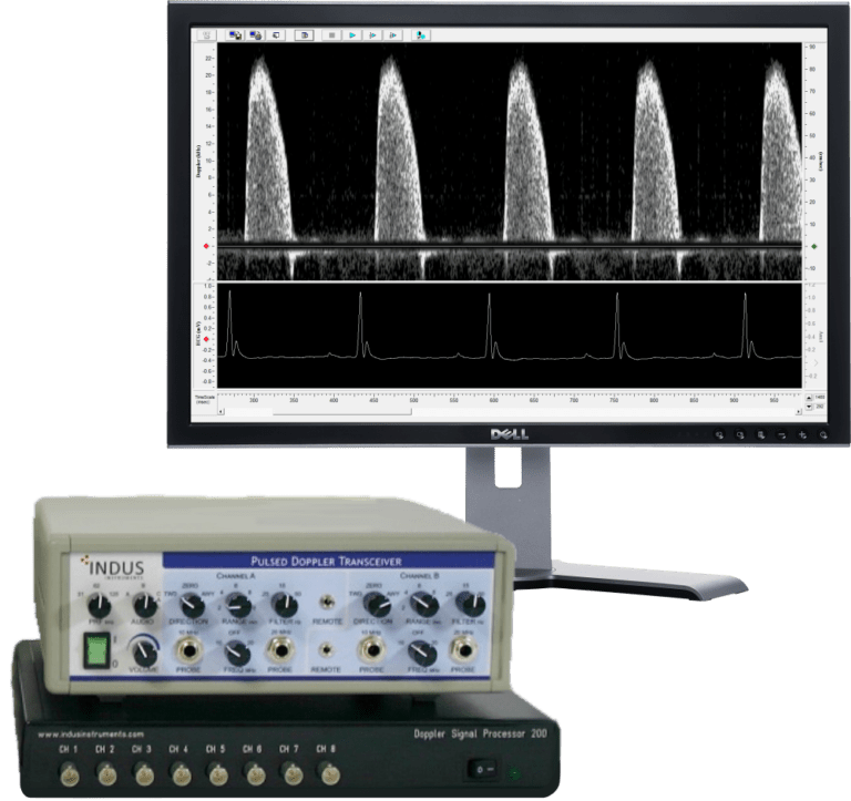
Doppler Flow
Velocity System
A Non-invasive Measure of Cardiovascular Physiology and Function
"Reliable and easy to use rodent surgical system"
Weifeng Dong
Merck
"Reliable and useful platform for surgery and ECG phenotyping"
Christopher Ward
BCM
"Very satisfied with the system and support...
Excellent customer support, no only for installation but for any follow up questions."
Oscar Bermeo
Ohio State University
System Overview
The Indus Doppler Flow Velocity System is a high-frequency, real-time pulsed Doppler measurement device with integrated data analysis software designed for measuring cardiovascular function in small animals. The high sampling rates grant excellent temporal resolution, making this the ideal system for studying fast heart rates and rapid blood accelerations present in small animals. Hardware components include the Pulsed Doppler Transceiver (PDT – a 10 & 20 MHz switchable dual channel system), Doppler Signal Digitizer (DSD), Doppler Workstation (DW), and handheld miniature probe(s).
Pulsed Doppler signals from the PDT are digitized at high sampling rates by the DSD and the workstation software. The acquired signals are processed using a fast Fourier transform algorithm and displayed as real-time grayscale Doppler flow velocity spectrograms. The workstation software permits recording and analysis of these spectrograms, ideal for report generation and publication purposes. This system has been successfully used in publications with mice, rats, bats, naked mole rats and other small animals. It is also capable of measuring blood flow velocities in larger animals using our implanted extra-vascular Doppler cuff probes.
Non-Invasive
Using a miniature handheld probe, flow velocities and differentials in various arteries, including the aorta, are reliably measured by placing the tip of the probe at a sharp angle relative to the direction of flow to be measured
Small Footprint
A powerful system that is both compact and easily transportable, allowing it to be scaled in larger facilities with ease or shared between cooperating labs without complication
Translational Data
Large bodies of literature support the translational relevance of rodent flow velocity data to clinical findings: study areas include cardiac function, myocardial perfusion, pressure overload, arterial stiffness, and more
Applications
Cardiac Function:
Systolic and Diastolic
Area
- Myocardial Infarction
- Heart Failure
- Hypertrophy
- Cardiomyopathy
Flow Parameter
- Aortic Outflow Velocity
- Mitral Inflow Velocity
Coronary
Flow Reserve
Area
- Myocardial Ischemia
- Pressure Overload-Hypertrophy
- Artherosclerosis
Flow Parameter
- Hyperemic/Baseline Coronary Flow Velocity Ratio
Arterial Stiffness
(Pulse Wave Velocity)
Area
- Hypertension
- Artherosclerosis
Flow Parameter
- Aortic Arch Velocity
- Abdominal Aortic Velocity
Pressure-Overload
(Stenosis)
Area
- TAC Banding Model
Flow Parameter
- Carotid (R/L) peak velocity ratio
- Stenotic jet velocity-estimation of pressure gradient across stenosis
Peripheral Artery
Disease and Perfusion
Area
- Renal, Carotid, Iliac, Femoral and Saphenous Vein Flow Velocities
Flow Parameter
- Flow Velocities in peripheral vessels before & after a surgical intervention or during therapeutic response
Parameters
Surgical Monitoring
& Vital Sign Measurements
- Heart Rate
- R-R Interval
Peripheral Artery:
Carotid, Renal,
Femoral & Tail
- Peak Velocity
- Mean & Minimum flow velocity
- Pulsatility Index
- Resistivity Index
Diastolic: Mitral
Inflow Velocity
- E-peak & E-stroke velocity
- E-time duration
- E-acceleration & E-deceleration time
- E-peak to ½ E-peak time
- E-linear deceleration time & rate
- A-stroke distance
- A-time duration
- E-A peak velocity ratio
- Isovolumic contraction time
- Isovolumic relaxation time
Other: Coronary, Transverse &
Abdominal Aorta
- Peak Diastolic Velocity (Coronary)
- Peak Systolic Velocity (Coronary)
- Diastolic & Systolic Area (Coronary)
- Ratios PSV/PDV & SA/DA
- Pulse Wave Velocity
Hardware
Engineered for precision, the transceiver delivers seamless transmit-and –receive functionality, while the digitizer transforms returning echoes into crisp, high-resolution digital data offering more reliable insights into flow dynamics.
Doppler Signal Digitizer
-
Channels – Channels 1 and 2 = Doppler InPhase & Quadrature
Channel 3 = ECG Channels 4 – 8 = Auxiliary inputs -
Input Range – ±10 V
-
Coupling – AC or DC software selectable
-
Sampling – 125 kHz per channel, 16 bits
-
Low Pass Filter – 10, 20, 30, 40, 50, 60, 70, 80, 90, 100, 110, 120, 130, 140 or 150 kHz (via Hardware)
-
Low Pass Filter – 10, 20, 30, 40, 50, 60, 70, 80, 90, 100, 110, 120, 130, 140 or 150 kHz (via Hardware)
-
High Pass Filter – 100, 200, 400, 600, 800, 1000, 1500 or 2000 Hz (Second or Fourth order, via Software)
-
Digital Signal Processor – 500MHz Dual Core Processor
-
Data Link to PC – USB 2.0 (480 Mb/s)
-
Power – 100-240 VAC Universal Adapter
Pulsed Doppler Transciever
-
Channels – Two, each switchable between 10MHz & 20MHz
-
Power – 110 VAC / 60Hz OR 220 VAC / 50Hz
-
Recorder Outputs – 2 from each channel (Phasic & Mean)
-
Audio Outputs – 2 from each channel (InPhase & Quadrature)
-
Audio Monitor – Amplifier & speaker selectable from any channel
-
External Ground – Intended for chasis grounding, if required
-
USB, RF/DEMOD – Future use
-
Transmitter Pulse Width – 0.4 μs
-
Receiver Pulse Width – 0.32 μs
-
Variable Range Gate – 1-10 mm (1-13 μs)
-
Velocity Outputs – 0.25 V/kHz simultaneous Phasic & Mean
-
Phasic Output Filter – Phasic (1 pole at 50 Hz), Damped (1 pole at 15 Hz) and Mean (2 poles at 0.25 Hz)
-
Probe Connection – Floating & differential (single-ended, differential)
-
Velocity Range – 1-100 cm/s at 0° angle, 2-200 cm/s at 60° angle
-
Electrical Zero – Front panel switches
-
Controls – Range adjustment, Polarity Switch, Filter
-
Ultrasound Frequency – 10MHz // 20MHz
-
Pulse Repetition Frequency – 31.25, 62.5, 125 KHz // 62.5, 125 KHz
-
Transmitter Output – 25 Vpp into 50 Ohm // 35 Vpp into 50 Ohm
-
Audio Bandwidth – ≈ 100 Hz to 15 KHz // ≈ 200 Hz to 25 KHz
Software
The software is designed to streamline your workflow with intuitive controls that makes it easy to analyze datasets. Designed with simplicity in mind, it provides the essential tools you need to manage analysis with reliable results.
Compatible Physiological Platform
The Rodent Surgical Monitor (RSM+) combines real-time, noninvasive physiological vital signs monitoring and surgical warming in a platform designed for mice, rats, and other similarly sized animals. Physiological signals can be directly integrated into the DFVS system for seamless data collection.
-
ECG
-
Respiration
-
Core Body Temperature
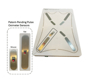
-
Oxygen Saturation
-
Ventricular and Systemic Pressure
High-Frequency Probes
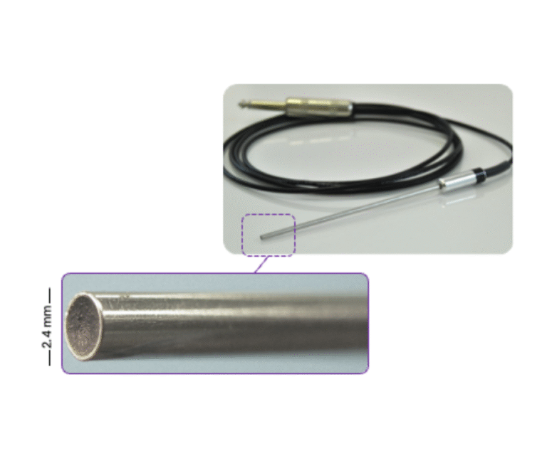
These probes are designed with high-quality piezoelectric crystals to provide superior spatial resolution for blood flow velocity measurements.
Available high-frequency probes:
- 10 MHz
- 20 MHz
- 30 MHz COMING SOON
-
Unique Pencil-like Design
~2 mm diameter probes allow for easier alignment to blood flow, which means more accurate data as compared to typical ultrasound probes.
-
Adaptability
Two probes can be used to image simultaneously at two different sites of interest. Taking variability out of applications like pulse wave velocity.
-
Durable
These probes are designed with durability in mind and are built to withstand impacts normally seen within labs.
-
Can be Handheld or Mounted with Micromanipulator
For long-term data collection, micromanipulators can be utilized to hold the probe stably
Doppler Transducers
Doppler transducers provide accurate flow velocity measurements in applications where handheld probes are not practical. They are well-suited for:
Chronic and implantable studies
Intraoperative vascular assessment
Use in larger animal models
Engineering and industrial flow applications
The transducer cuff is constructed from flexible silicone and split longitudinally, allowing direct placement around the vessel for secure and stable data acquisition.
Available Cuff Sizing
-
1.0 mm
-
1.3 mm
-
1.7 mm
-
2.2 mm
-
2.5 mm
-
2.8 mm
*Custom sizing is available
Imaging Gallery - Doppler Flow Velocity System
Get access to our exclusive video presentations
Due to the sensitive nature of the content, registration is required to get access to these resources. (Immediate Access)
Pulse Wave Wave Velocity
Coronary Flow
Transverse Aortic Constriction
Publications & Articles
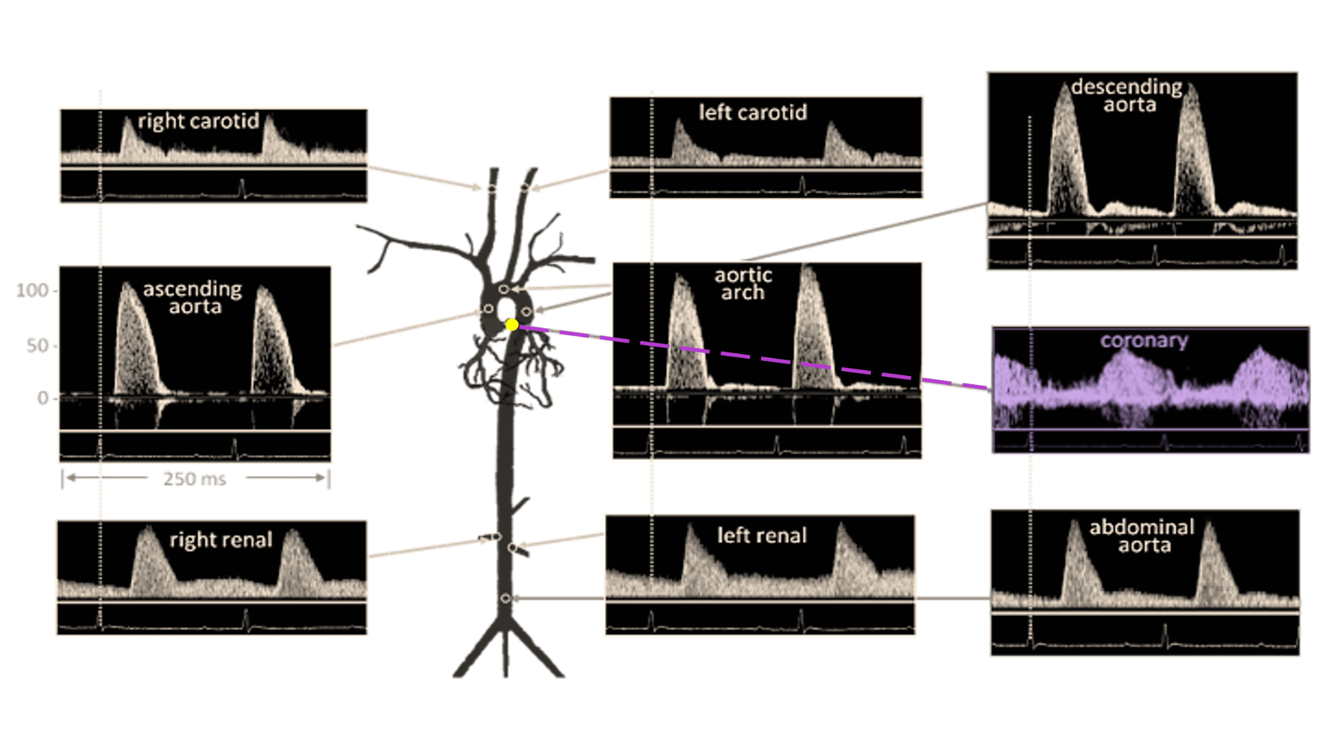
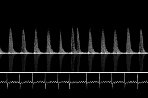
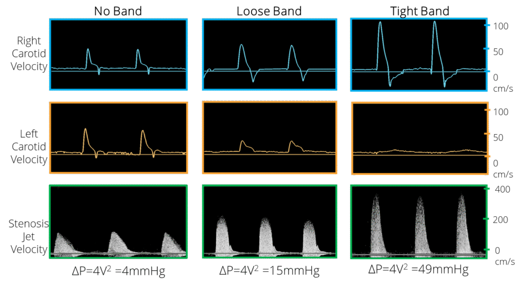
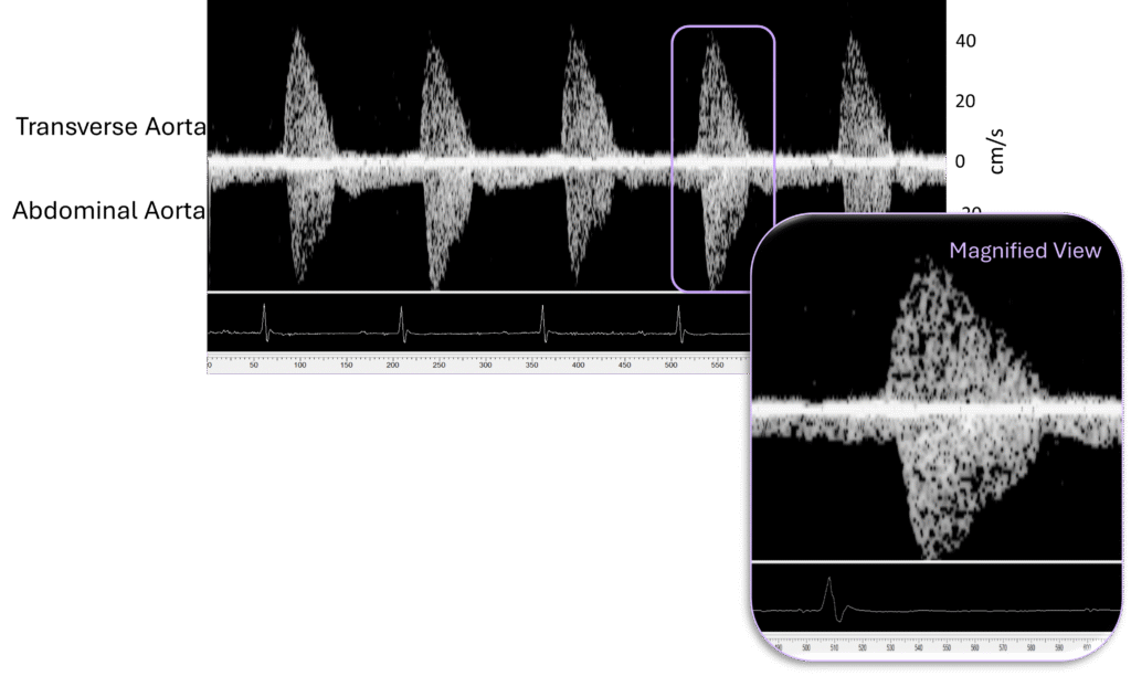
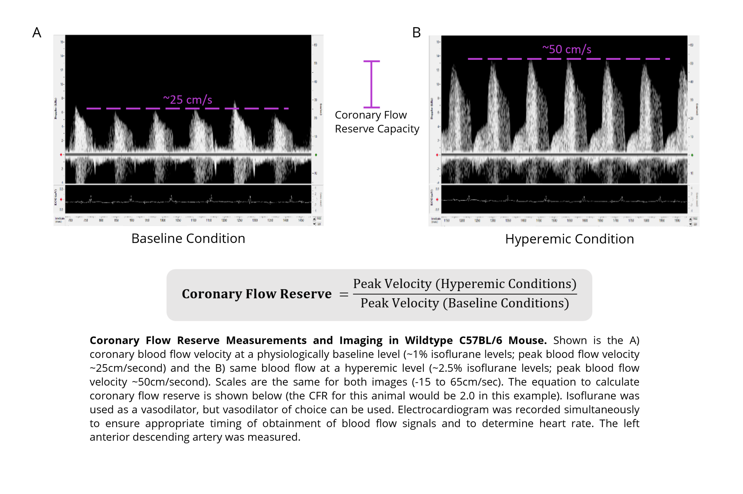
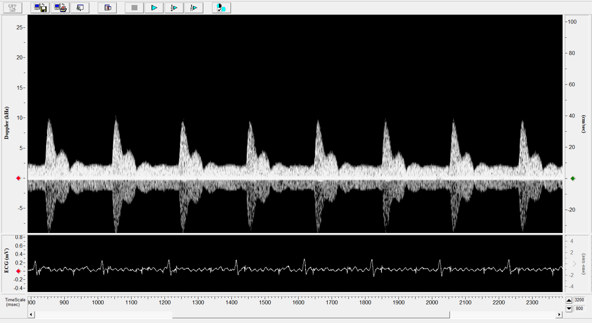
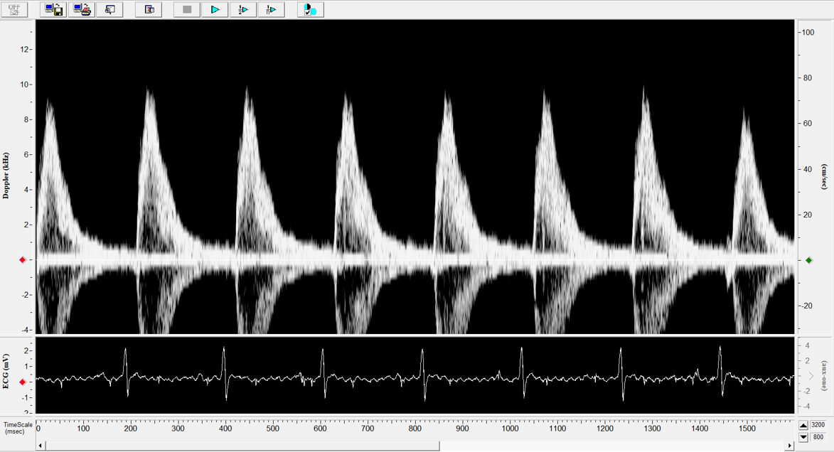
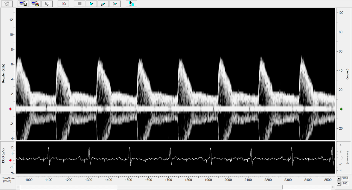
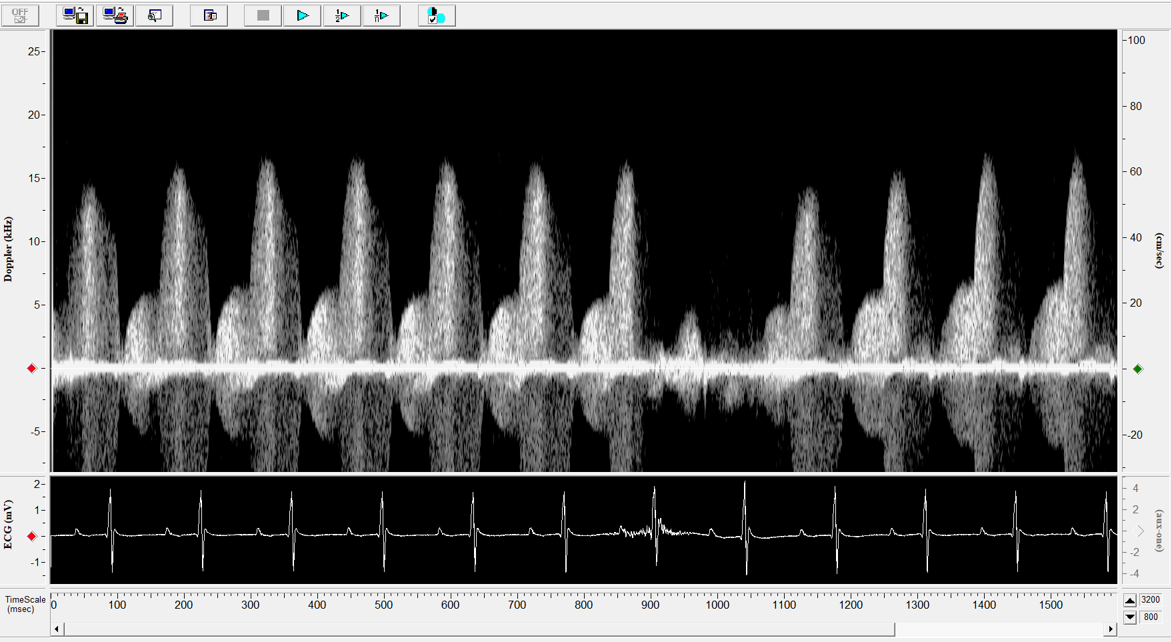
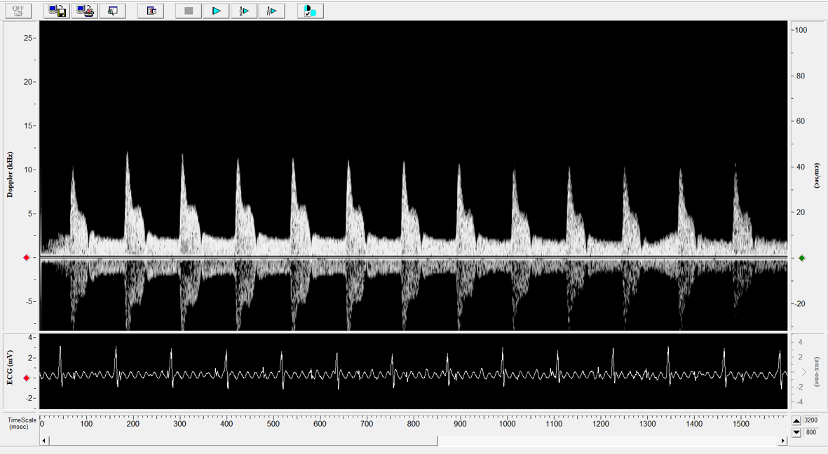
Application Note: Measuring Coronary Flow Reserve
System Used: Doppler Flow Velocity System M-Series View Paper Here Background Coronary flow
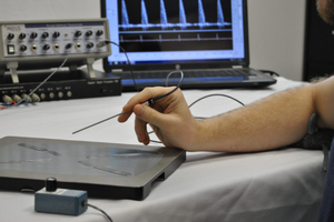
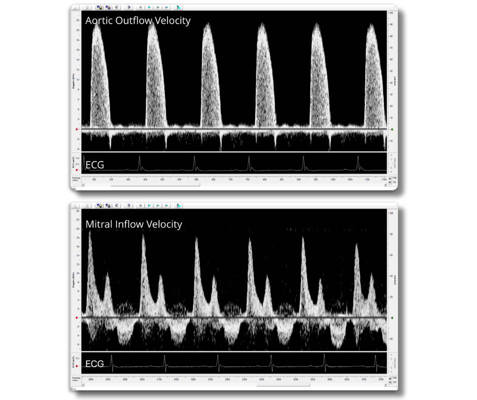
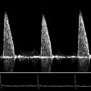
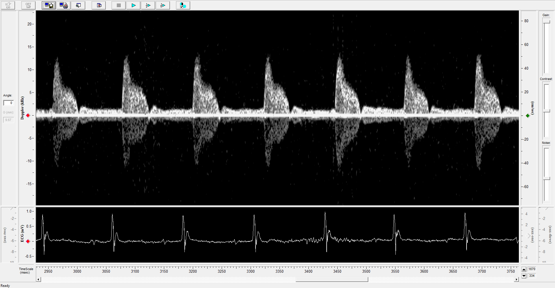
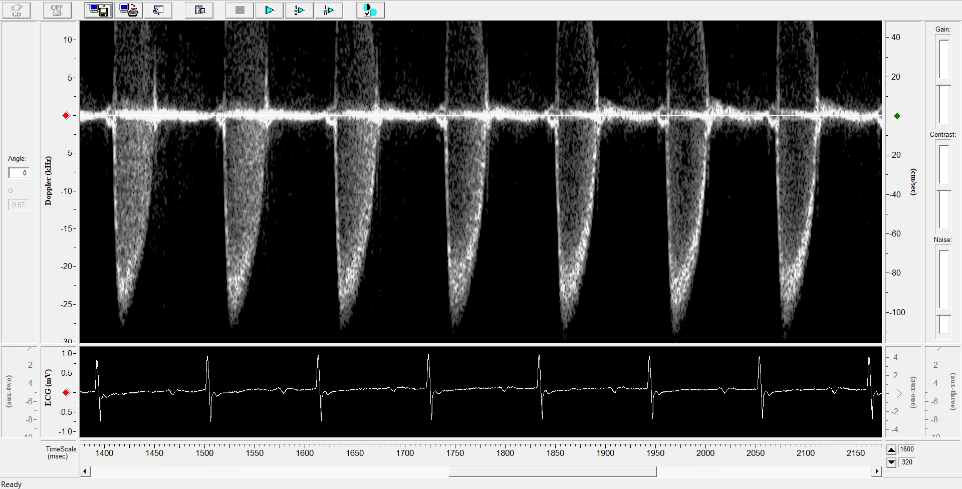
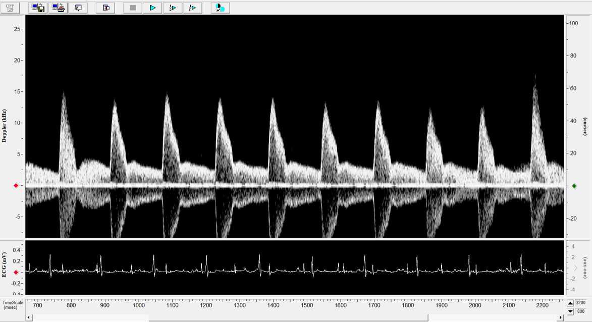
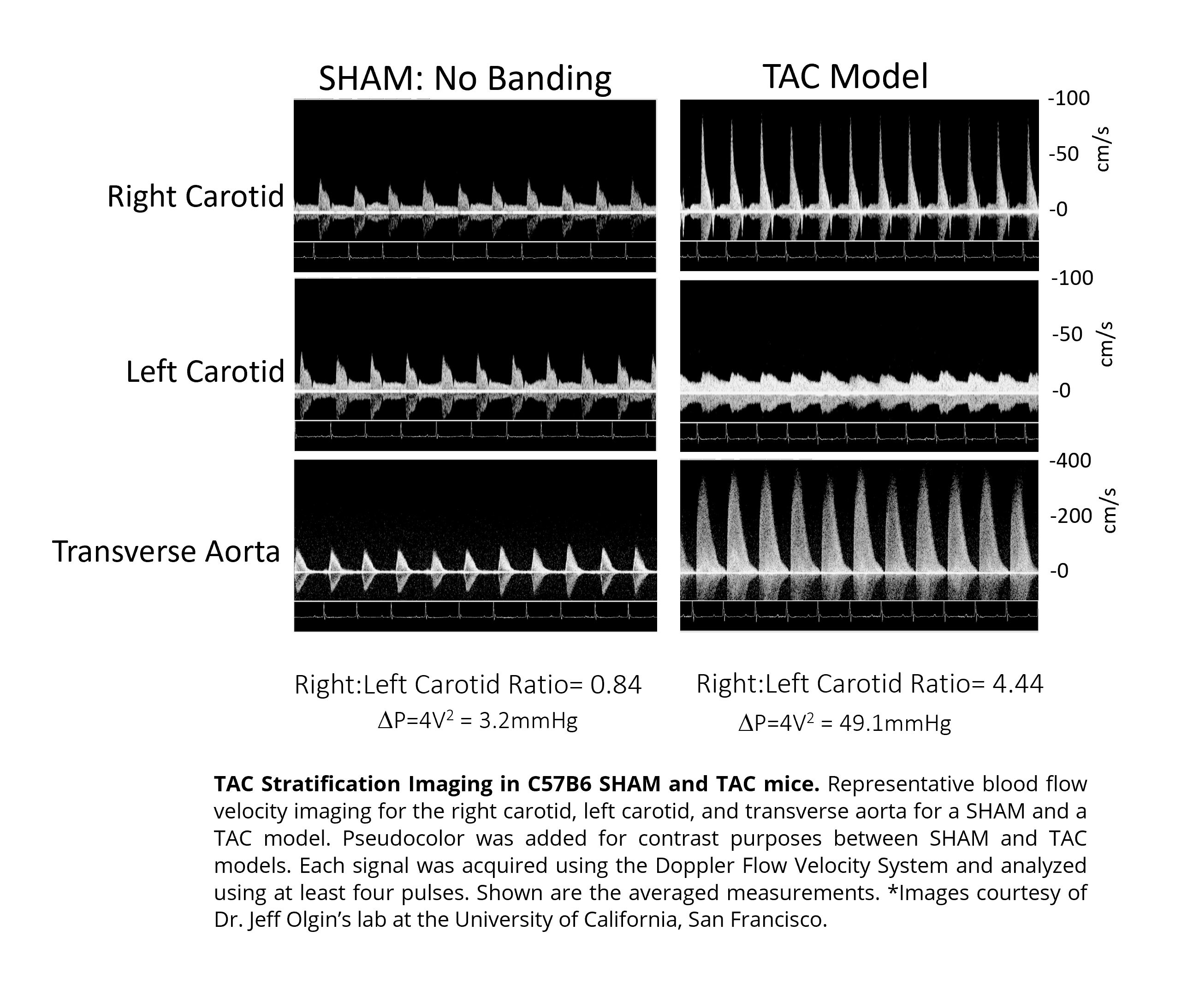
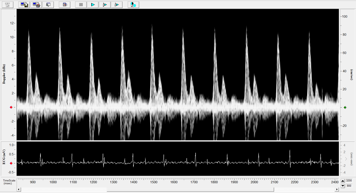
Science Corner: Doppler Flow Velocity Imaging at Dr. Olgin’s Research Lab
Doppler Flow Velocity Imaging at Dr. Olgin’s Research Lab Dr. Jeff Olgin. “Dr.
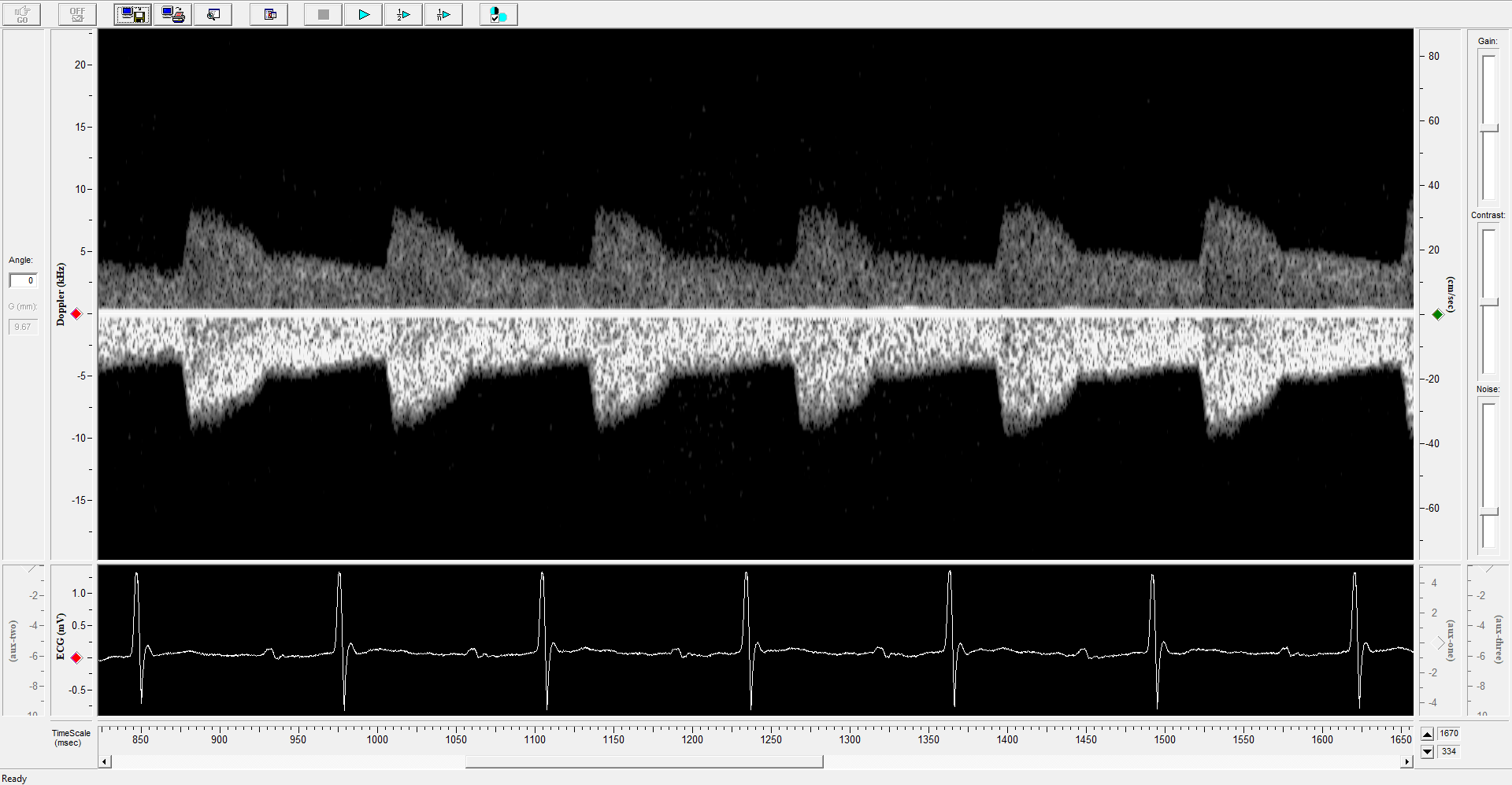
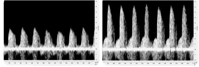
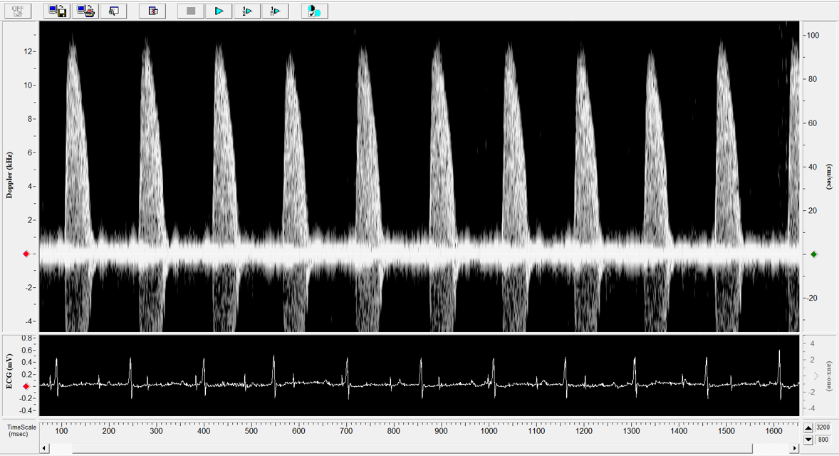
Article review: “Effects of Isoflurane on Coronary Blood Flow Velocity in Young, Old, and ApoE−/− Mice Measured by Doppler Ultrasound”
Article review: “Effects of Isoflurane on Coronary Blood Flow Velocity in Young, Old,