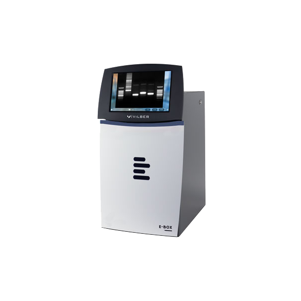
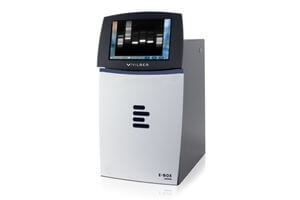
E-Box CX5
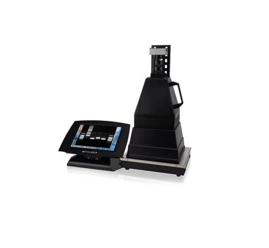
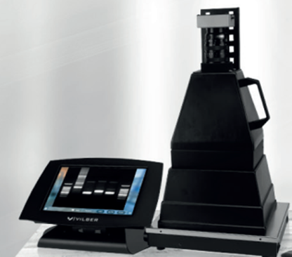
Vilber Doc-Print CX3
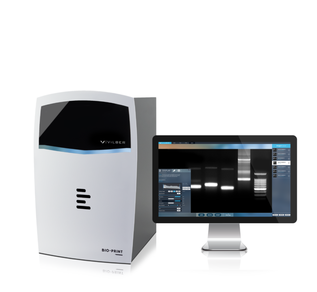
Vilber Bio Print CX4
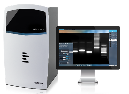
Vilber Quantum CX5
E-Box CX5
Vilber Doc-Print CX3
Vilber Bio Print CX4
Vilber Quantum CX5
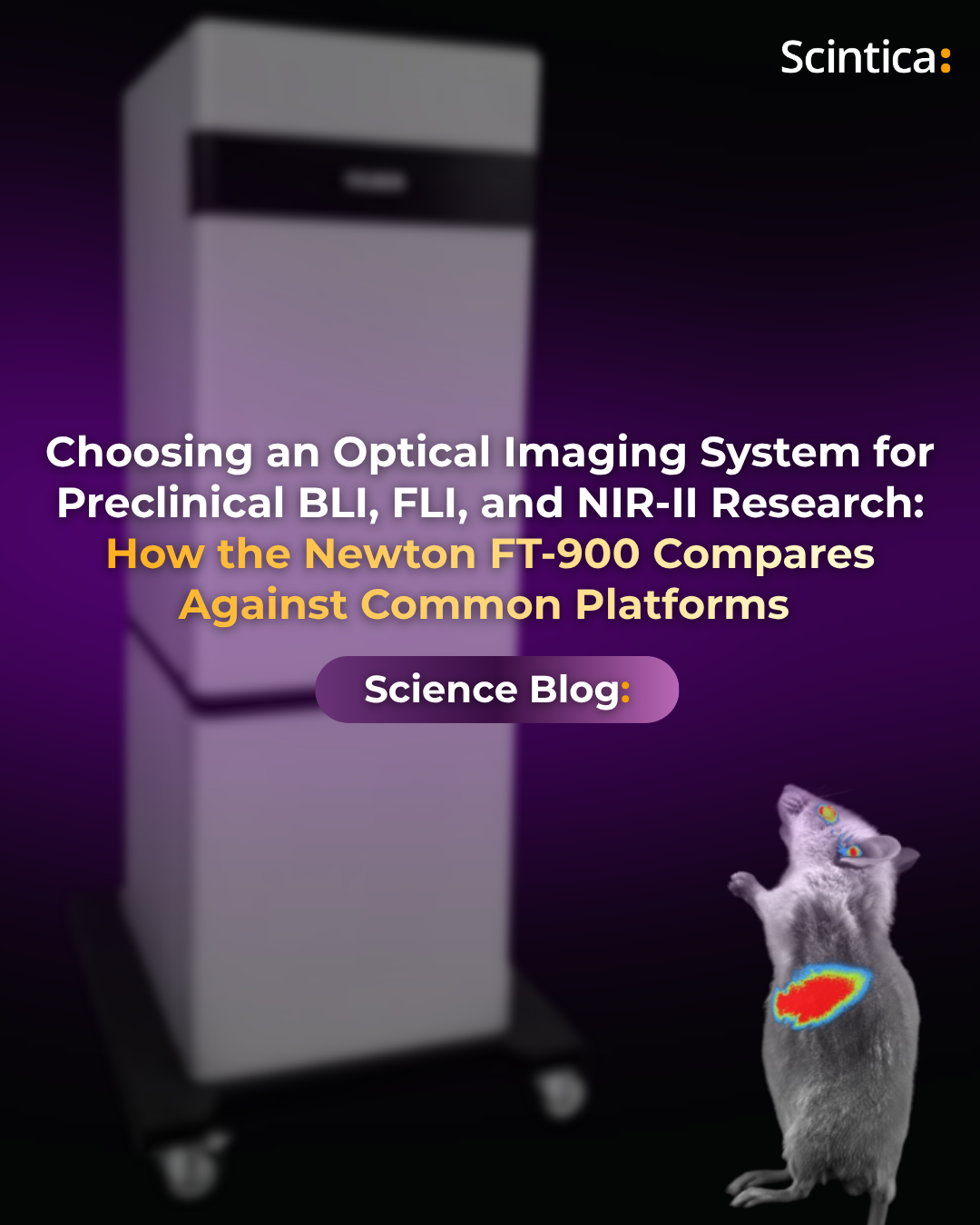
Choosing an Optical Imaging System for Preclinical BLI, FLI, and NIR-II Research
Choosing an Optical Imaging System for Preclinical BLI, FLI, and NIR-II Research: How
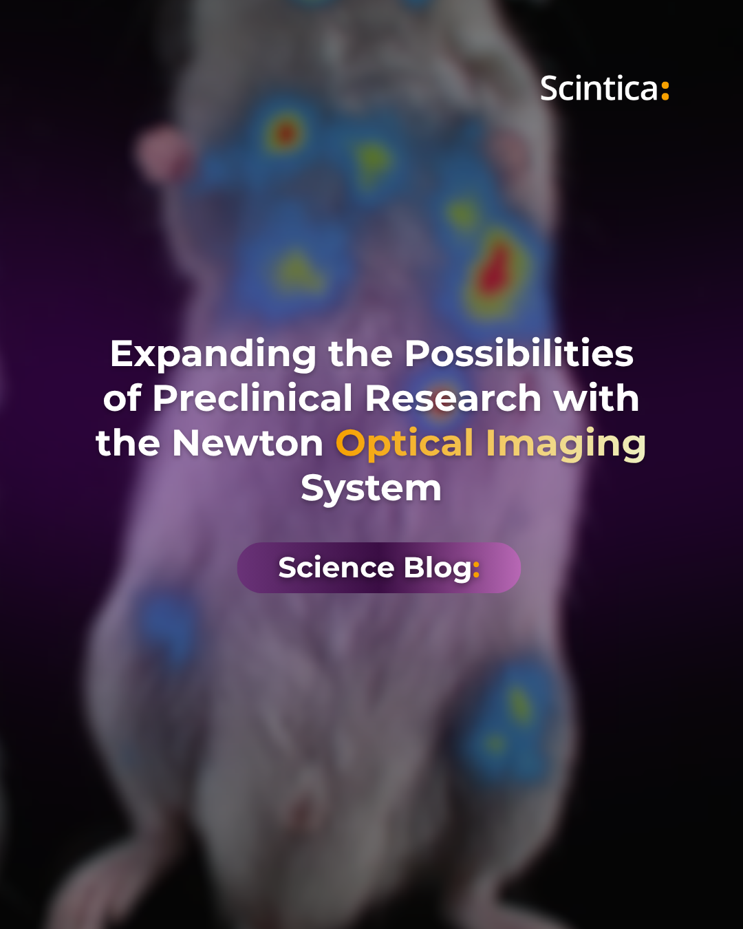
Expanding the Possibilities of Preclinical Research with the Newton Optical Imaging System
Expanding the Possibilities of Preclinical Research with the Newton Optical Imaging System Understanding
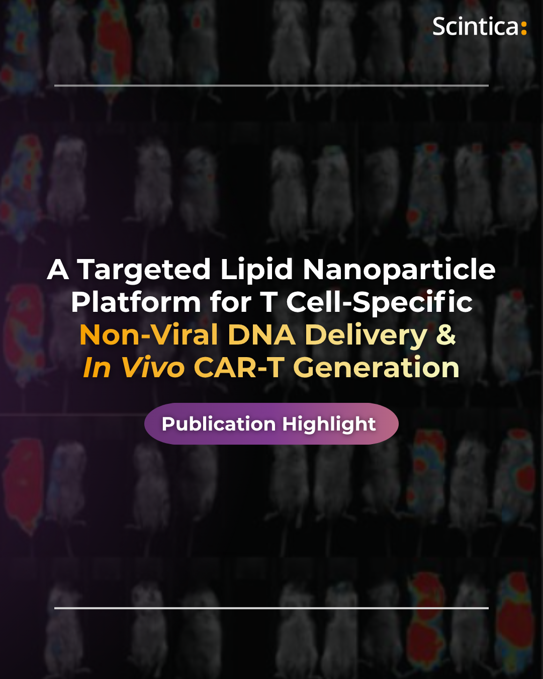
Publication Highlight: T Cell-specific Non-viral DNA Delivery & In Vivo CAR-T Generation Using Targeted Lipid Nanoparticles
Publication Highlight: A Targeted Lipid Nanoparticle Platform for T Cell-Specific Non-Viral DNA Delivery
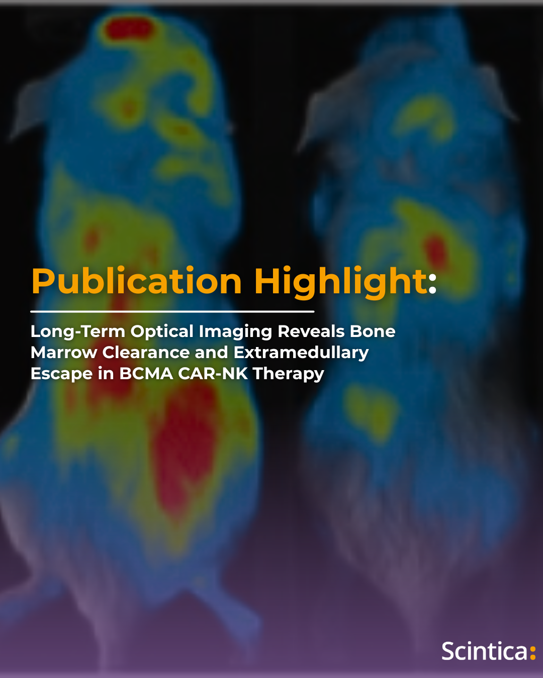
Publication Highlight: Long-Term Optical Imaging Reveals Bone Marrow Clearance and Extramedullary Escape in BCMA CAR-NK Therapy
Publication Highlight: Long-Term Optical Imaging Reveals Bone Marrow Clearance and Extramedullary Escape in
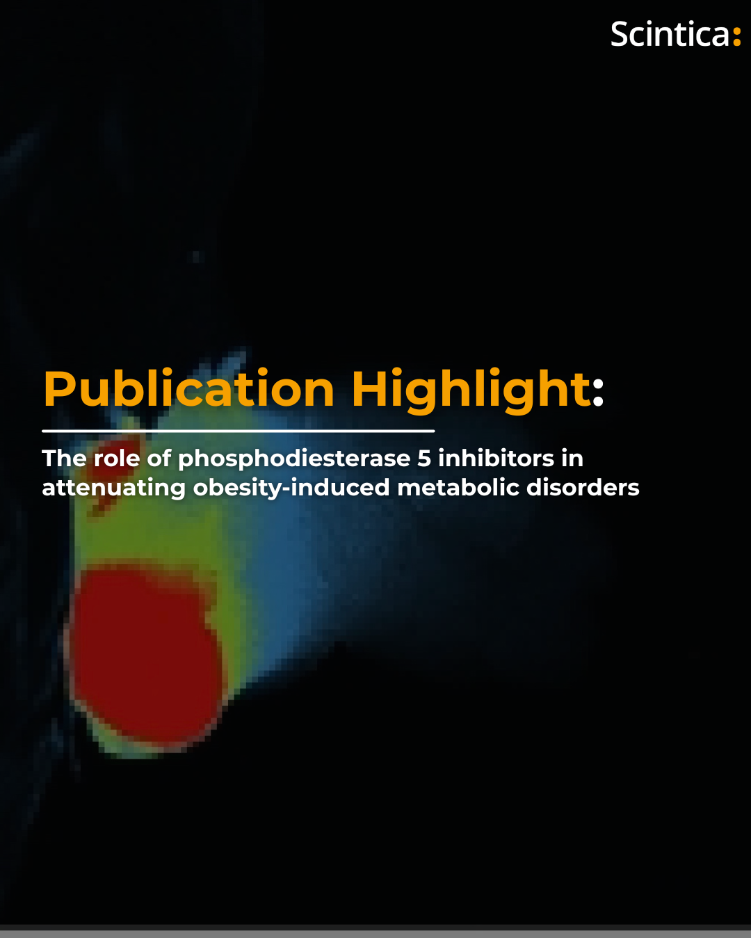
Publication Highlight: The role of phosphodiesterase 5 inhibitors in attenuating obesity-induced metabolic disorders
Publication Highlight: The role of phosphodiesterase 5 inhibitors in attenuating obesity-induced metabolic disorders
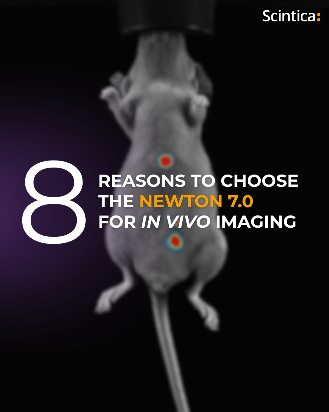
8 Reasons to Choose the Newton 7.0 for In Vivo Imaging
The Newton 7.0 sets a new benchmark in preclinical optical imaging. Designed for reliability and versatility,