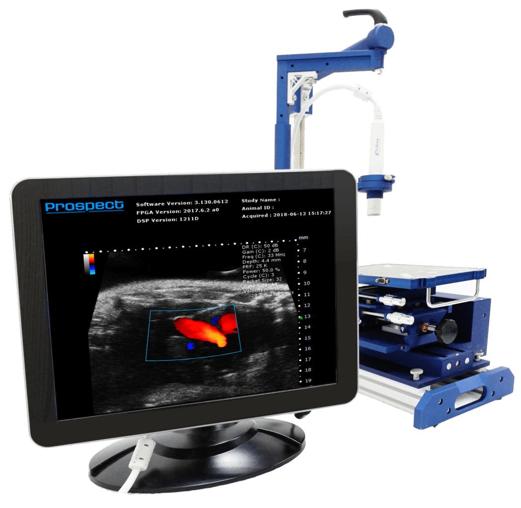
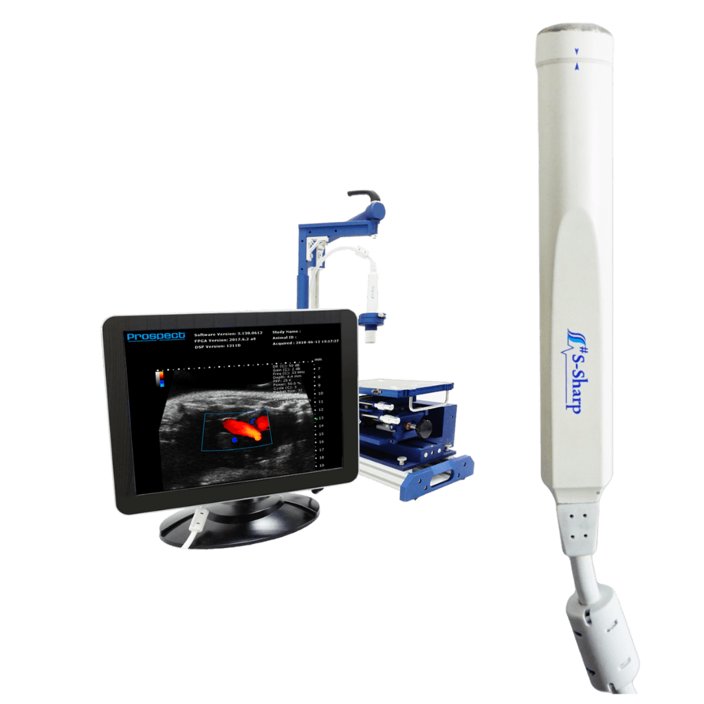
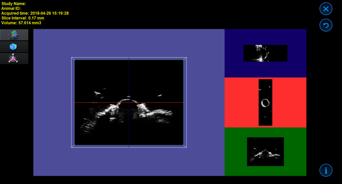
3D Motor and Acquisition/Analysis Software
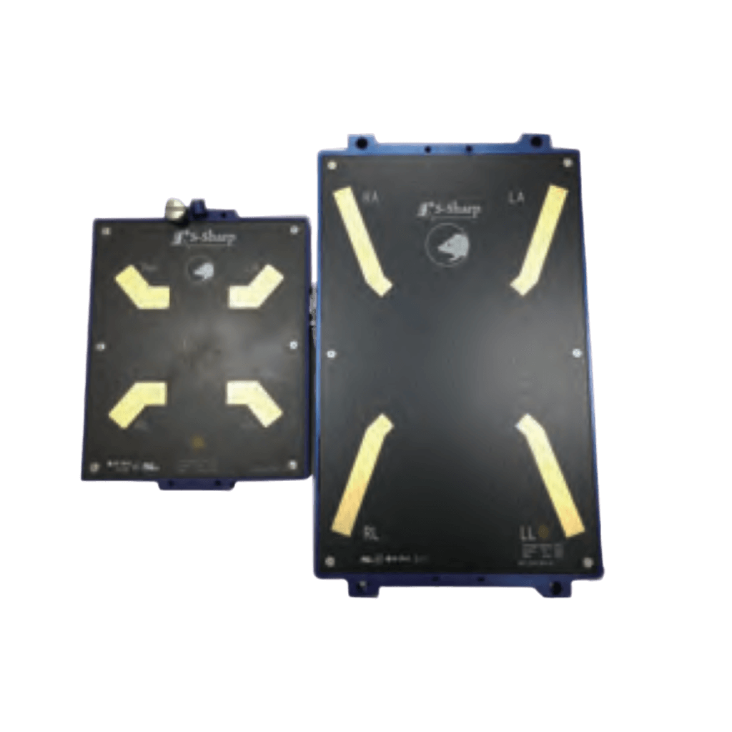
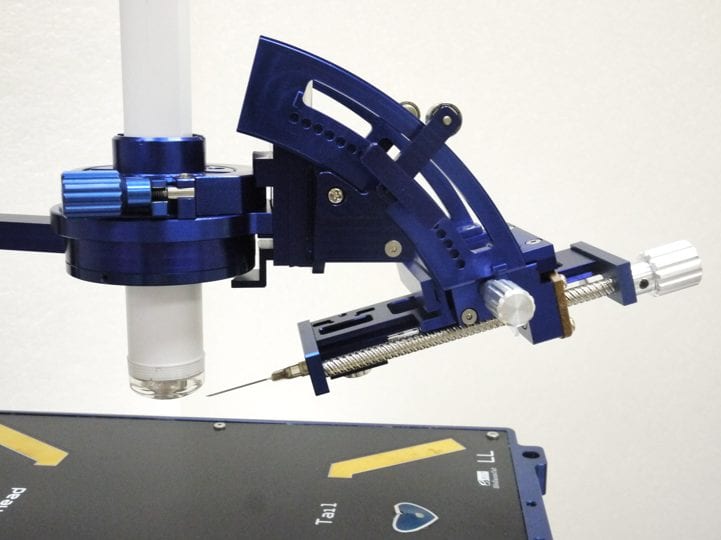
Image-guided Needle Injection Mount
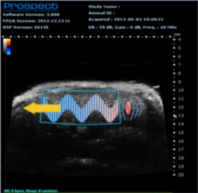
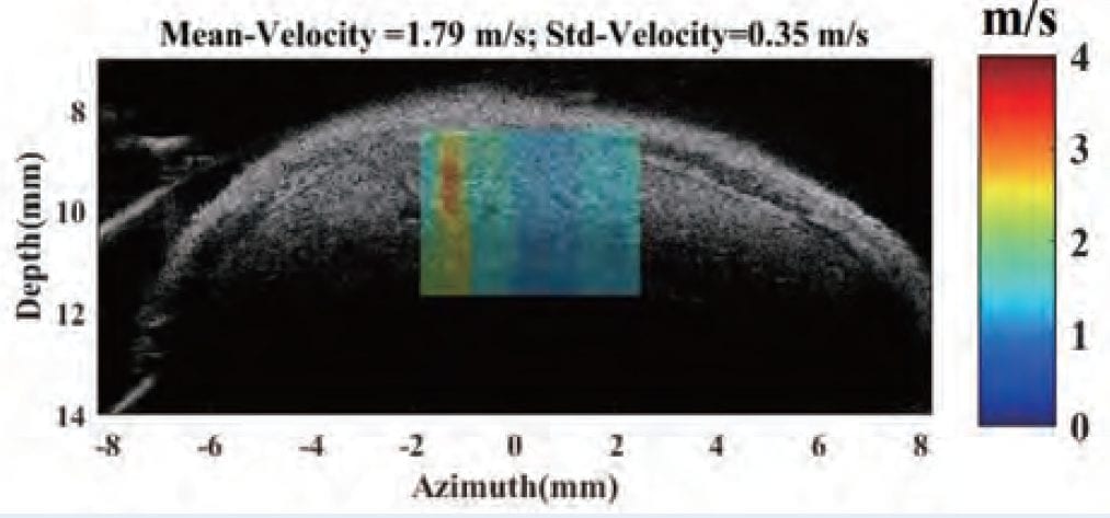
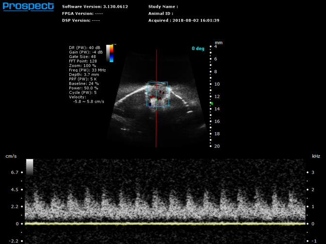
Integrated Shear Wave Elastography Probe and Analysis Software
3D Motor and Acquisition/Analysis Software
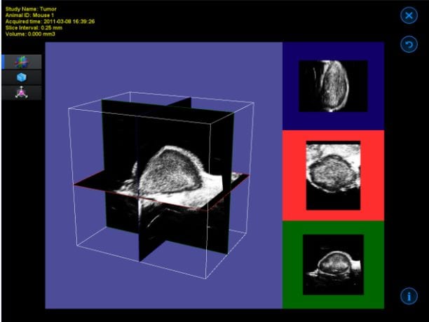
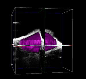
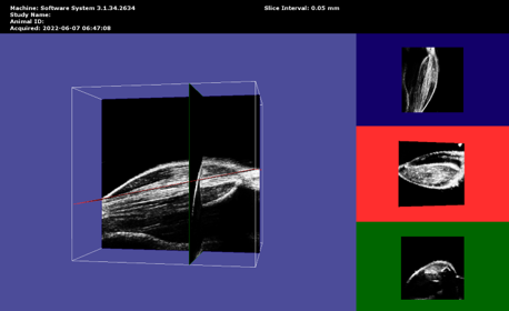
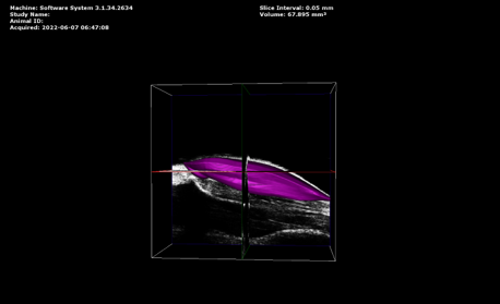
Image-guided Needle Injection Mount
Integrated Shear Wave Elastography Probe and Analysis Software
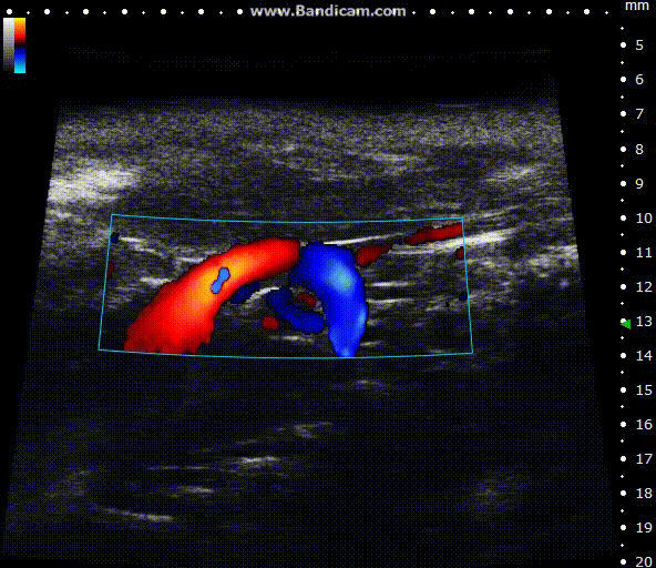
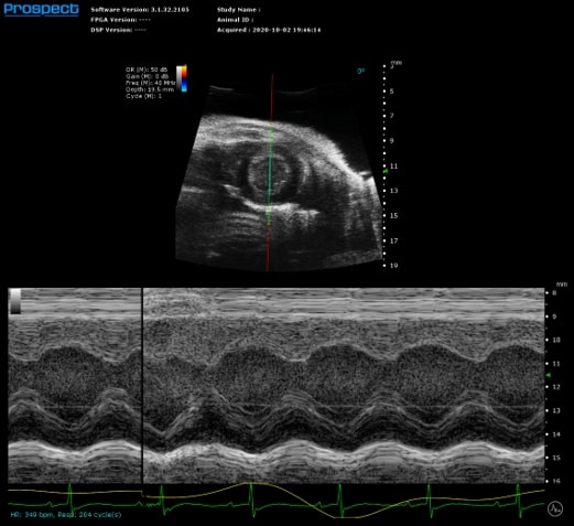
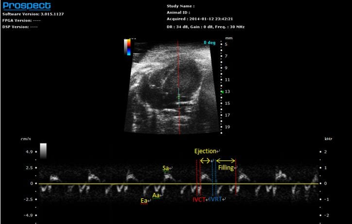
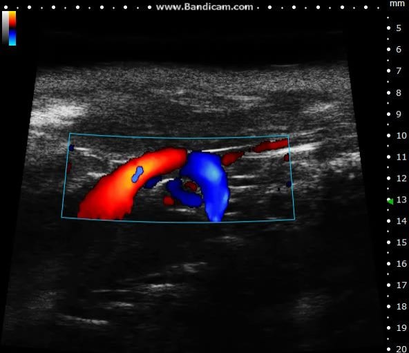
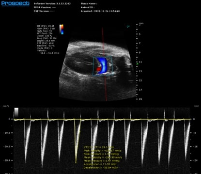
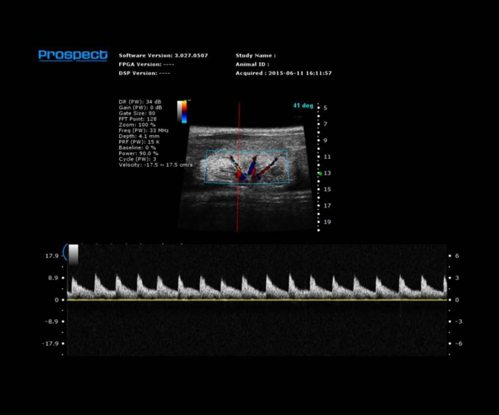
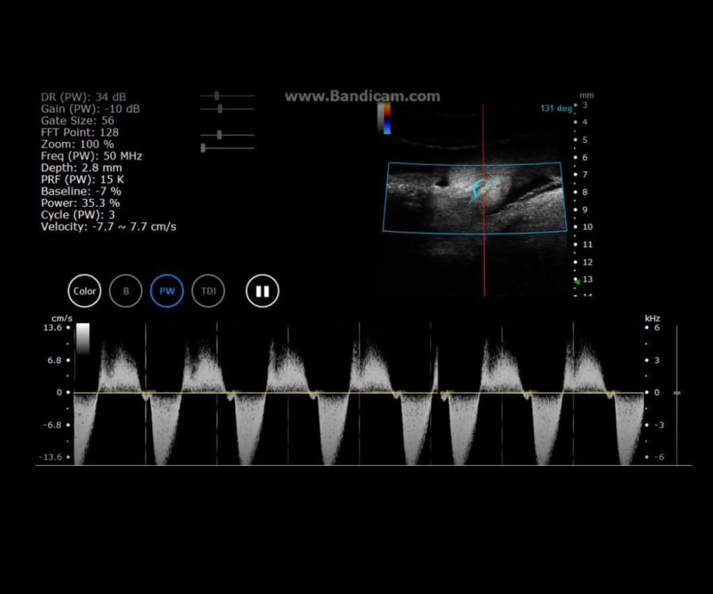
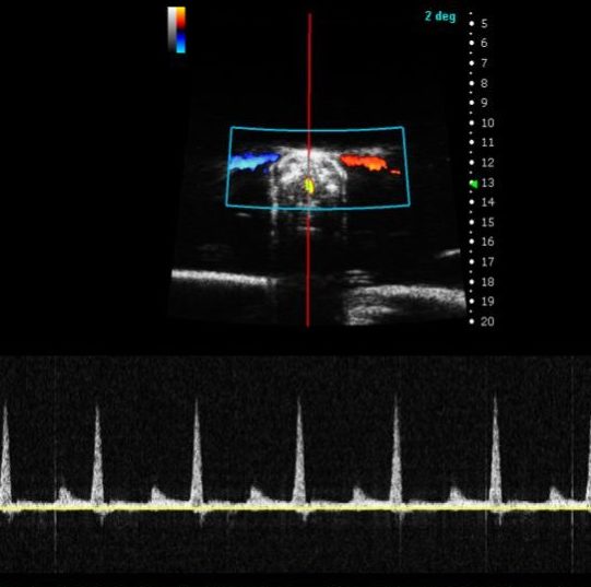
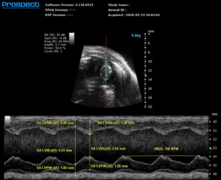
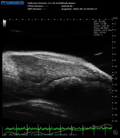
Cardiovascular Biology
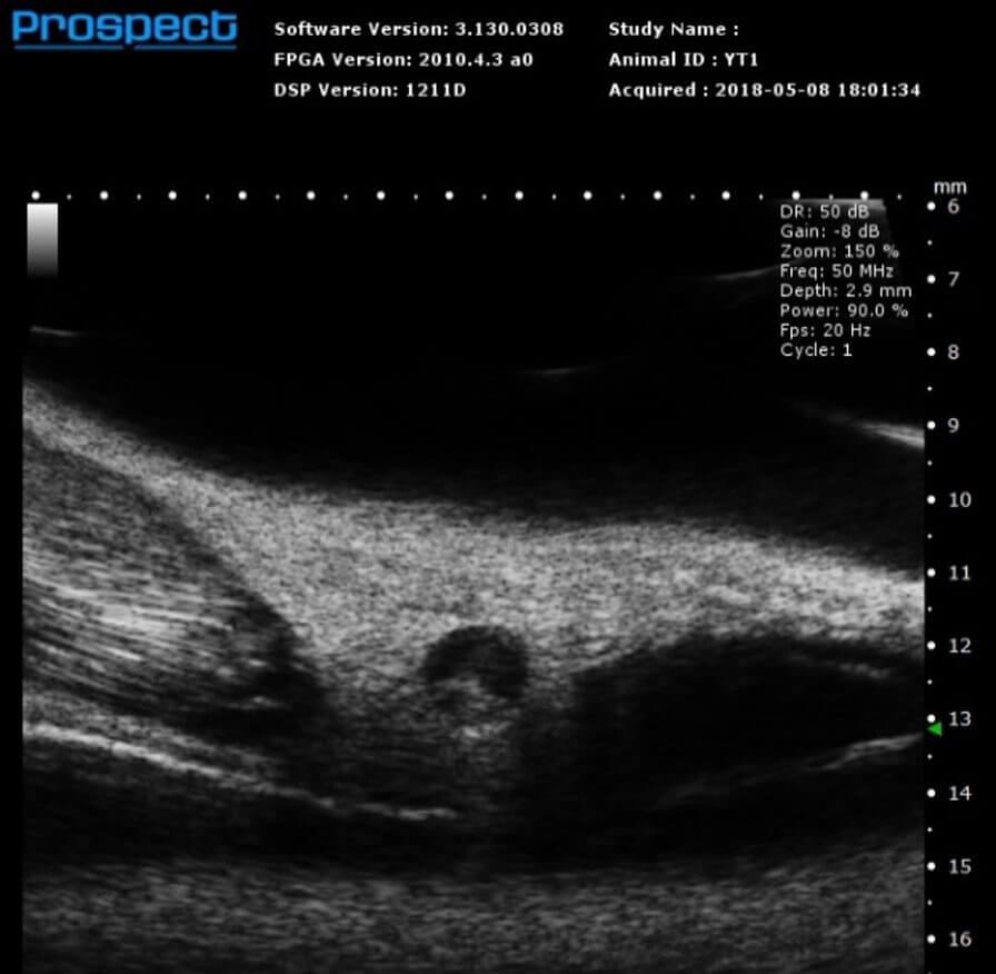
Cancer Biology
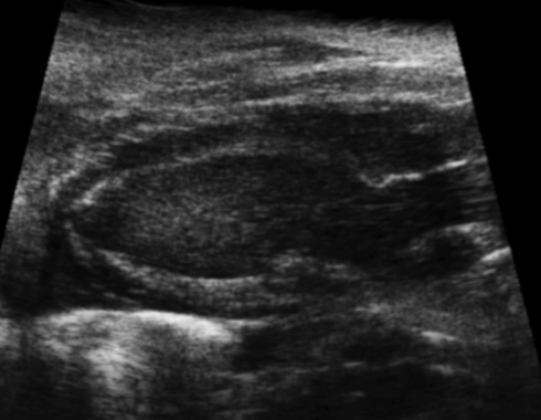
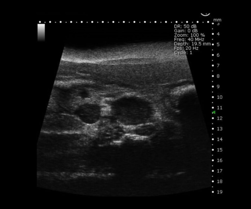
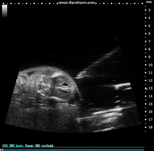
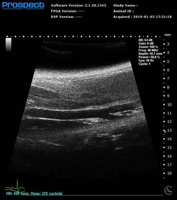
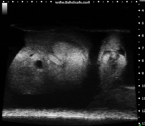
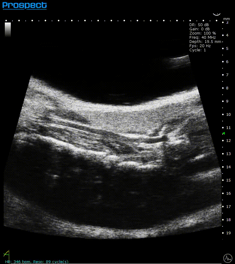
Abdominal & Anatomical Imaging
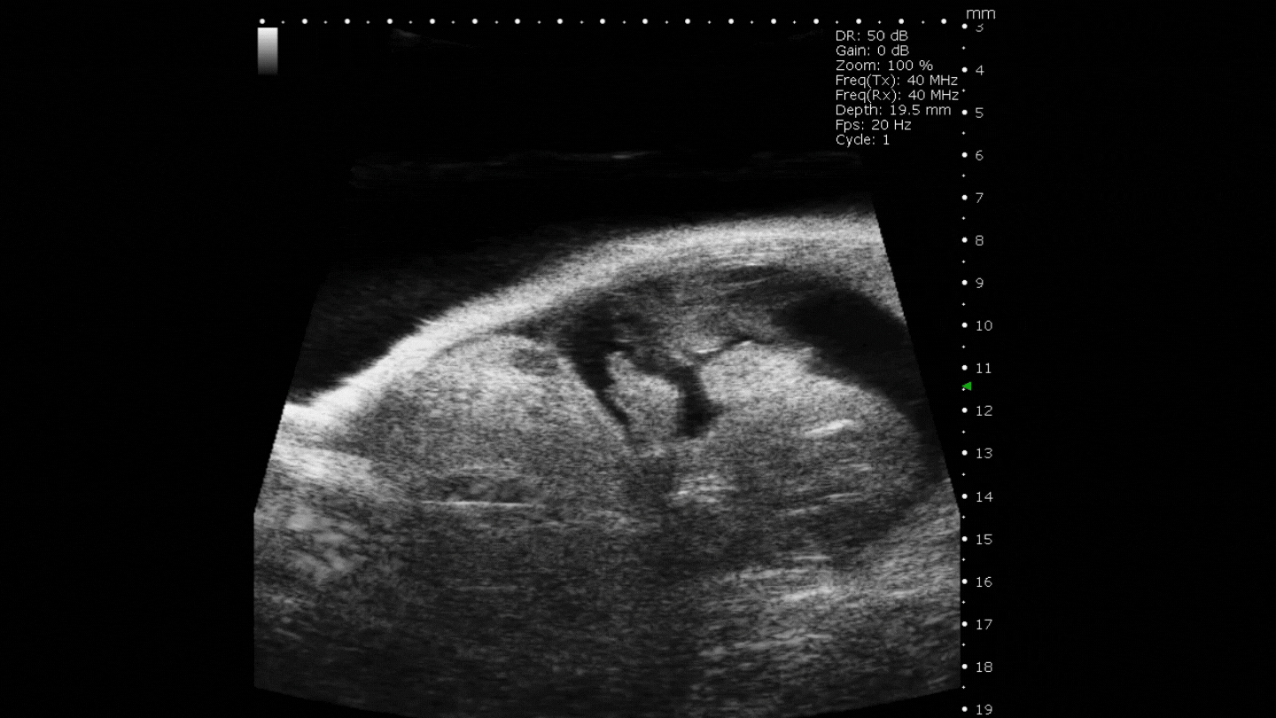
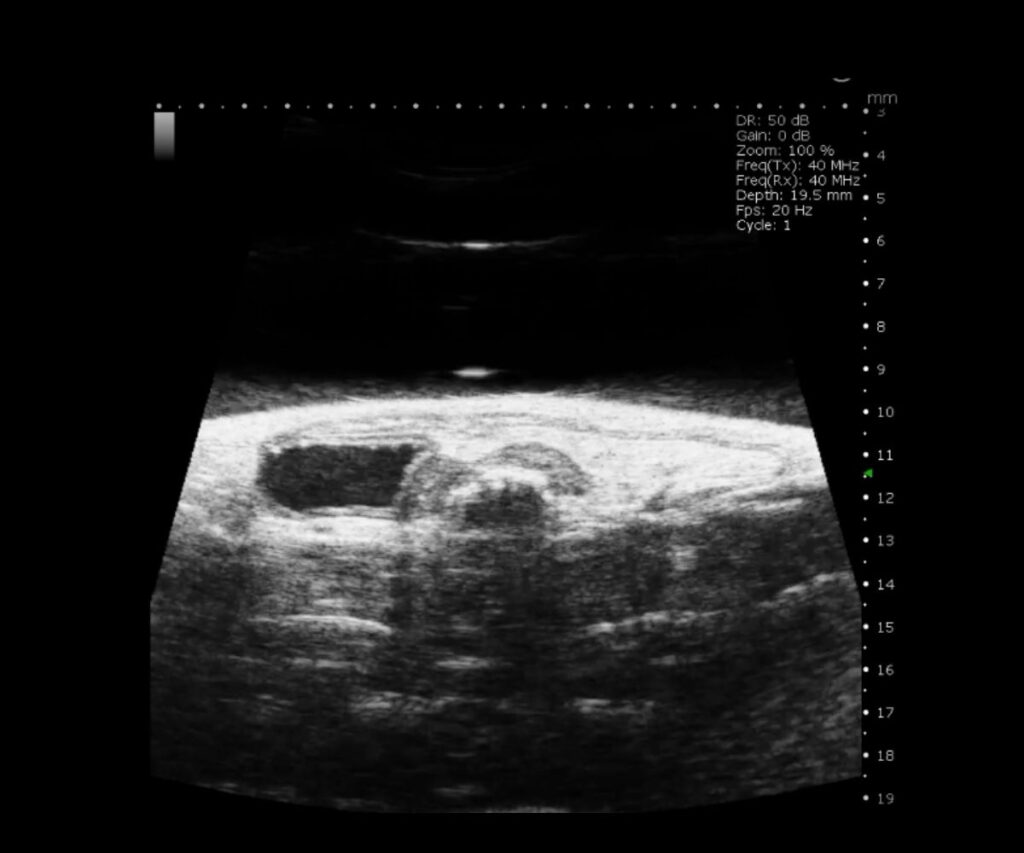
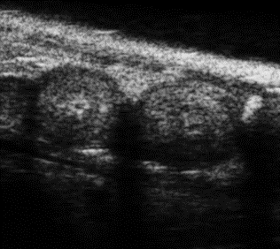
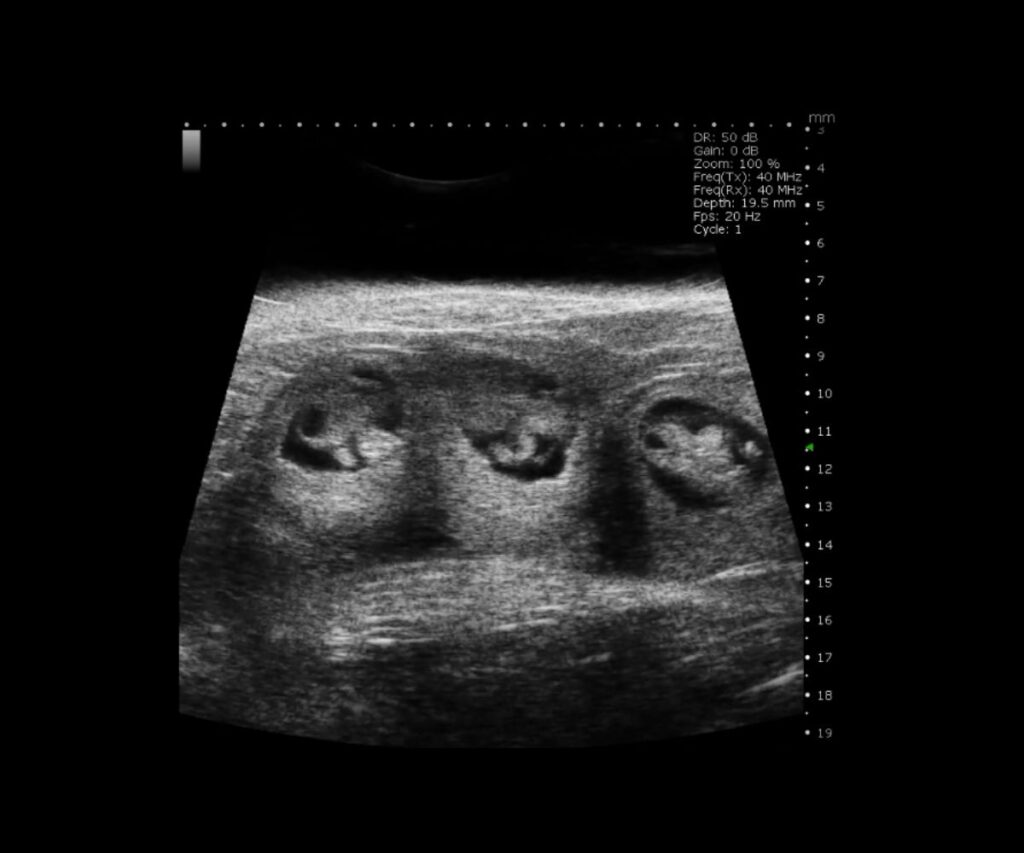
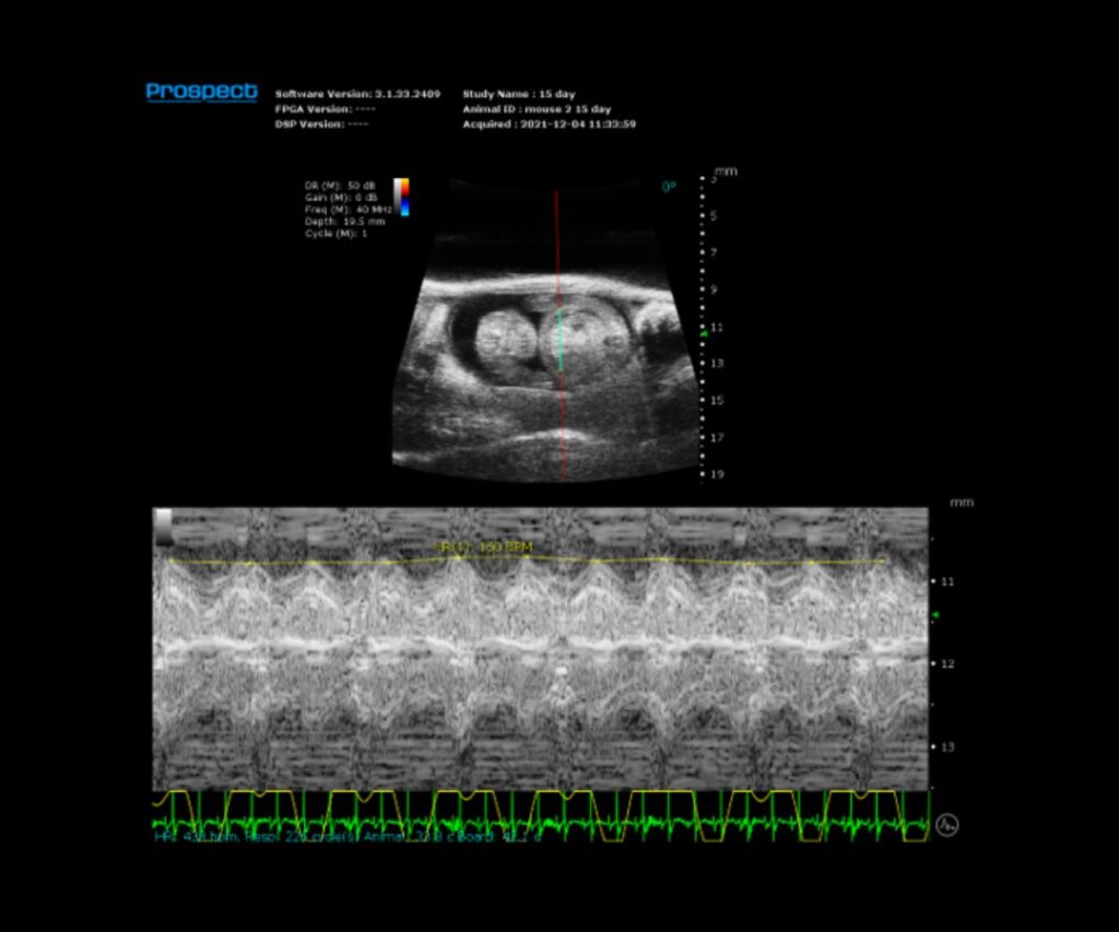
Developmental Biology
Ophthalmology
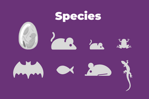
Other Species
Cardiovascular Biology


Cancer Biology
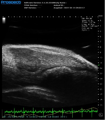
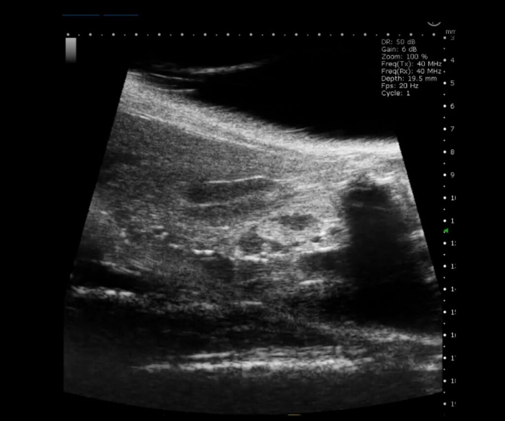
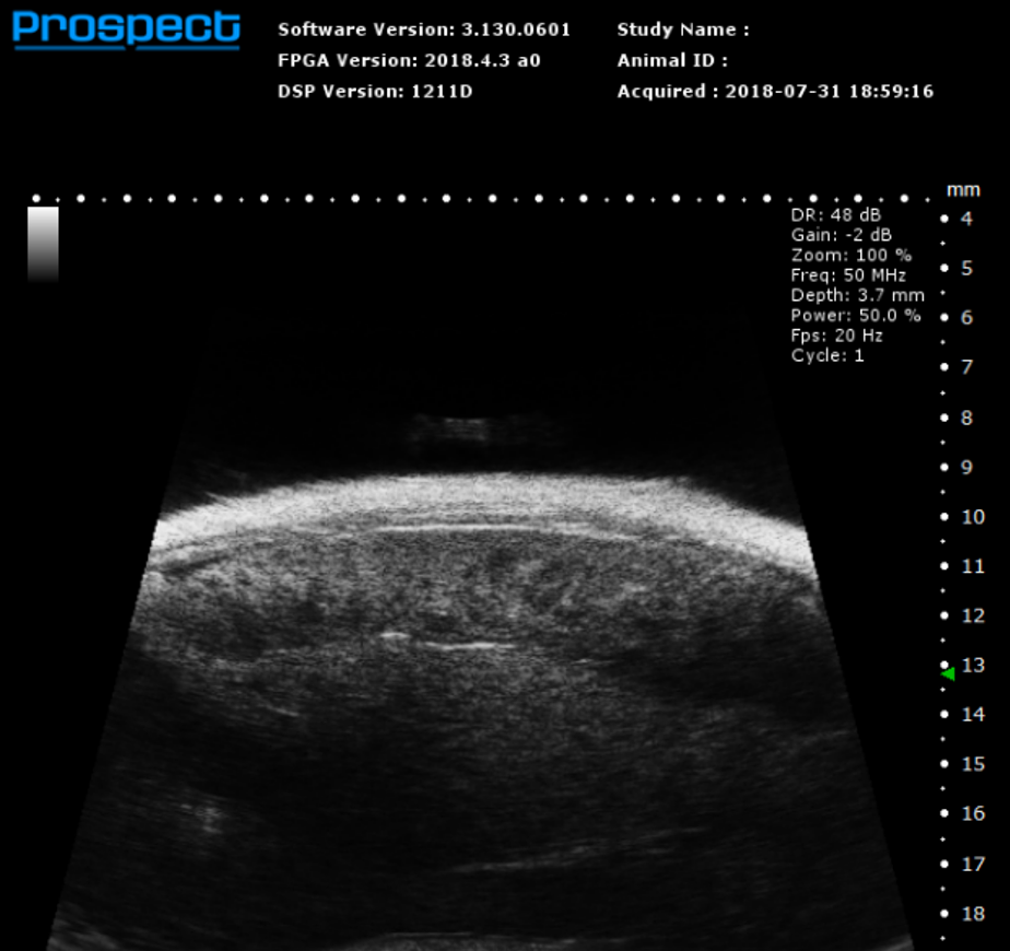
Abdominal & Anatomical Imaging
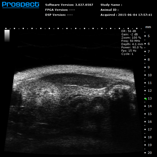
Developmental Biology
Ophthalmology
Other Species

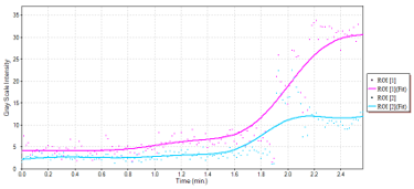
Science Corner: Innovations in Radiolabelling Techniques
Innovations in Radiolabelling Techniques and Radioimmunotherapy Enhanced by Ultrasound Imaging Dr. Jason Holland’s
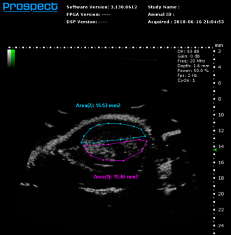
Allogeneic Expanded Human Peripheral NK Cells Control Prostate Cancer Growth in a Preclinical Mouse Model of Castration-Resistant Prostate Cancer
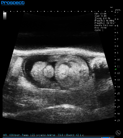
System Used:Prospect T1 Fangming Wang, Xuejiao Dong, Jing Wang, Feiya Yang, Donghua Liu,
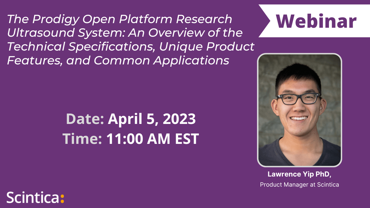
(April 5, 2023) Webinar: Prodigy Open-Platform Research Ultrasound System Overview
(April 5, 2023) Webinar: Prodigy Open-Platform Research Ultrasound System Overview Overview:In this webinar,
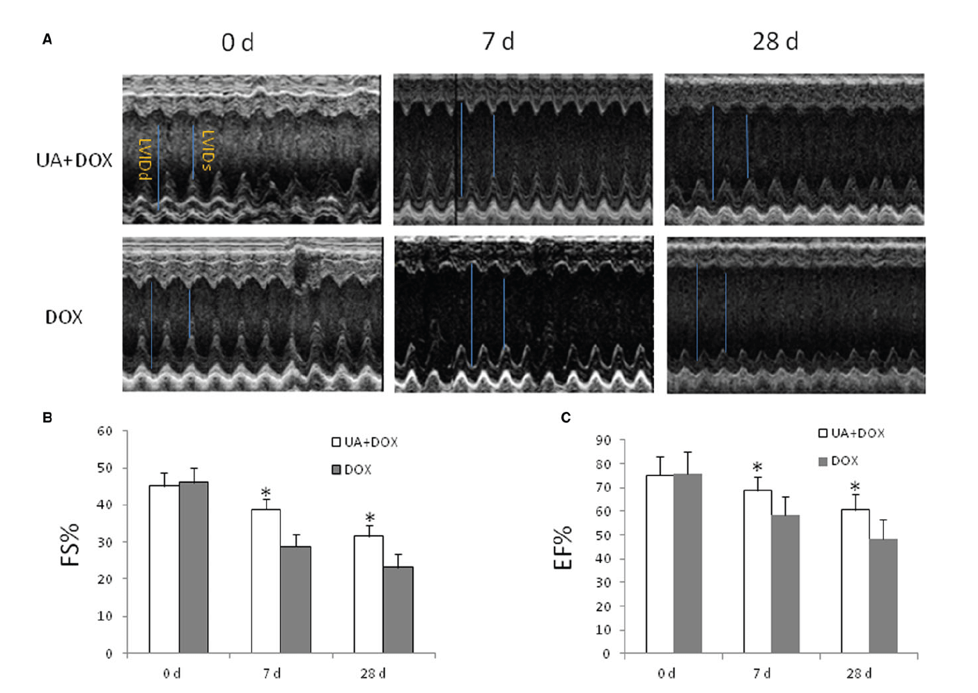
Ursolic acid prevents doxorubicin‐induced cardiac toxicity
Ursolic acid prevents doxorubicin‐induced cardiac toxicity in mice through eNOS activation and inhibition
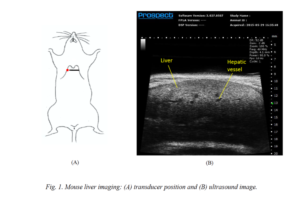
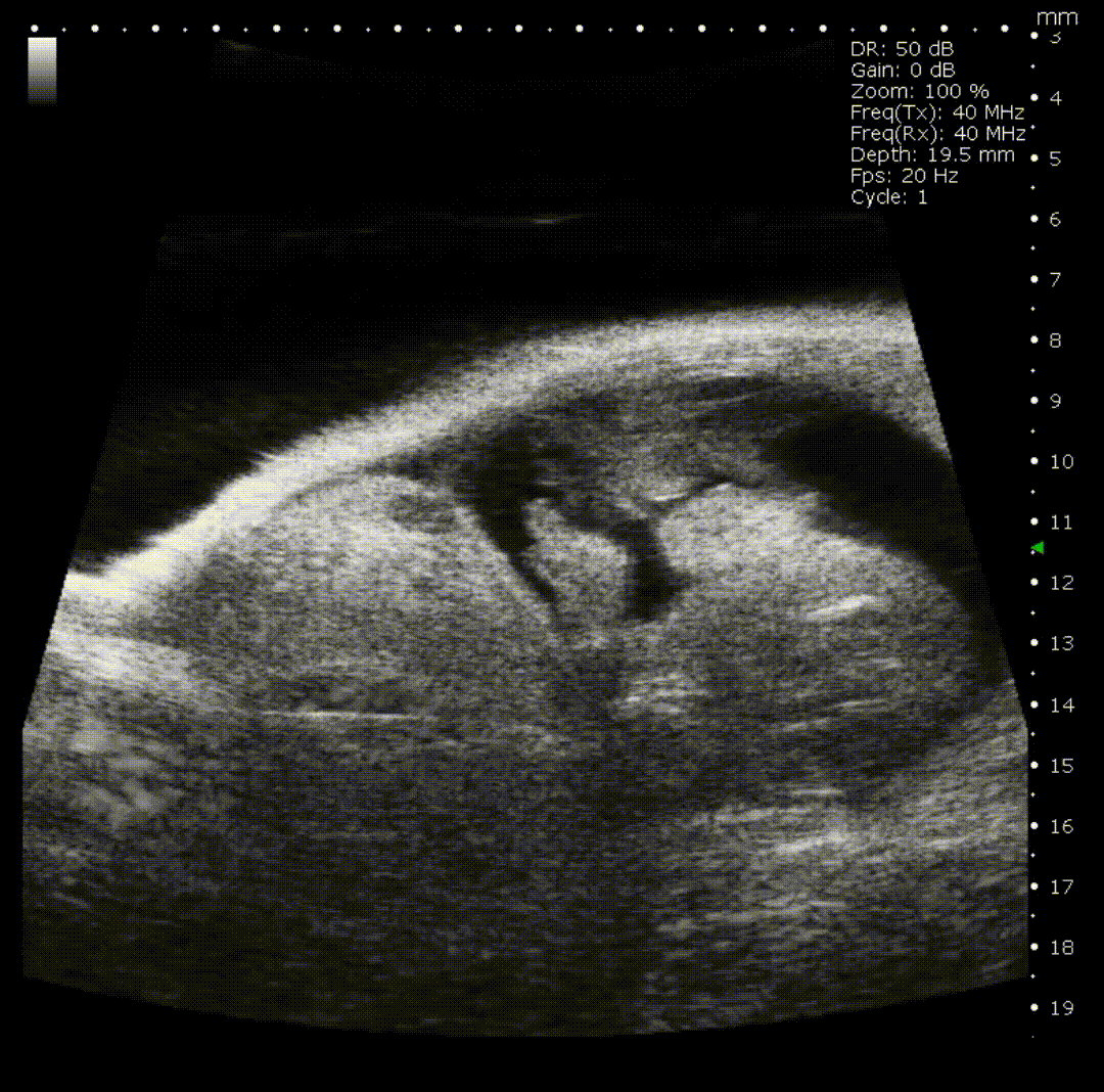
The Mouse Abdominal Imaging Guide
Mouse Abdominal Imaging Guide: Imaging the Liver, Hepatic Vessels, Portal Veins and Gallbladder
Applications in Developmental Biology
How the Prospect T1 high-frequency ultrasound system can be used in preclinical developmental