What is live cell imaging and what sets it apart?
Live cell imaging is an umbrella term that refers to a plethora of methods that use time-lapse microscopy to observe, track, and quantify dynamic processes in cells or subcellular systems. This type of imaging is vital for a variety of fields including cell biology, developmental biology, neuroscience, biophysics, molecular biology, and cancer biology, to name a few.
While imaging of fixed cells can provide a useful snapshot of cellular behaviors, live-cell imaging removes the events that occur between data points and provides greater information about the dynamic processes which otherwise may be challenging to obtain. Additionally, there is no debate that information held within living cells is much more likely physiologically relevant to the understanding of cellular processes. Further to this point, fixation may even influence the way cells look, as different fixation methods can manipulate the apparent structure of the cytoskeleton or change cell morphology altogether.
Moving on, live cell imaging microscopes actually allow users to keep cells under well regulated environments. The simplest way to ensure cells are kept in controlled environments is by performing imaging directly within cell culture incubators (or workstations). This is achieved by placing a small microscope within the incubator and provides a direct method for imaging that diminishes environmental shock to cells. Of course, regular, manually controlled microscopes do not have this ability or functionality and do indeed lead to shocking cells even if the researcher is diligent.
COVID-19 is changing how labs function, how can live cell imaging help?
While not every institution or lab has the same physical distancing or occupancy restrictions, nearly every research group will have to adapt in some way to the new world we live in. Because of the novel Coronavirus, many labs are being forced to scale back research as lab personnel are often required to work in shifts. These labs, therefore, are only able to get a fraction of the work they would normally accomplish done now. In this sense, being able to automate some simple tasks or assays may be key in keeping many research objectives on schedule.
Live cell imaging microscopes have a real advantage moving forward as they can allow for researchers at home (or anywhere with an internet connection) to monitor cell growth dynamics. This opens up the lab for other researchers to do more work off-site and gives more hours in the day for other scheduled tasks. For instance, a researcher needing to do the simple task of checking confluence would no longer need to come into the lab but could rather leave the space open for their colleagues to get work done. On the same note, that same researcher could still come into the lab but not focus the precious time they have to a menial task, like checking confluence, until absolutely necessary. All in all, live cell imaging systems will make a lab more efficient in a time where efficiency is more essential than ever.
Beyond this, live cell imaging systems also provide additional information like growth curves or migration curves. This is also helpful for researchers today as it means not having to run a simple analysis which can be costly timewise. On that note, users do have access to the images and data and can run further analysis, as necessary.
Lastly, and extremely important to note is, culturing cells without live cell imaging is a black-box experiment. In between inspection rounds, assumptions are made of cell growth rates and cell viability based on the researcher’s experience. When using a live cell imager, these assumptions are no longer required as new images are automatically generated and can be assessed immediately. Researchers, without having to come into the lab, can update themselves on the health, migration, or growth dynamics of cells.

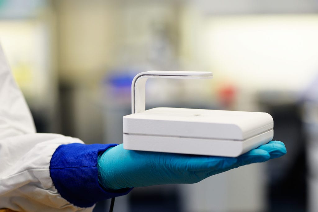
The CytoSMART Lux2 – A live cell imaging solution
Scintica Instrumentation offers the CytoSMART Lux2 as an easy and cost-effective solution for time-lapse imaging of cells. Users can monitor cell proliferation, motility, and morphology for hours or weeks at a time. The Lux2 is a compact inverted lab microscope for bright-field live cell imaging. It works from inside most incubators or workstations and can be used in routine cell culture processes. The Lux2 provides researchers with the ability to study morphology, migration, cytotoxicity, proliferation and more.
If you would like more information on the Lux2, visit the webpage here or reach out to sales@scintica.com and we’d be happy to help.