In my day to day interactions, I come across numerous researchers who are not able to fully utilize MRI systems as they would like or cannot see themselves using this technology even though it could immensely help their research. The reasons for this are numerous but often fall into 1 of 3 categories.
- Price – either that of the physical technology/running costs or to pay a core facility for use
- Too difficult – a background in MR physics is essentially needed to use most MRI systems
- Scheduling conflicts – the researcher is always on the schedule of a dedicated MR technician and the availability of the equipment within a core facility
I am here to suggest that an MRI system does exist that can remove these barriers and I fully intend to cover each of these points below. With that in mind, the only important question for you really is; can it work for your research and help you answer the biological questions you have? In the article below I will cover each of these questions as I review the Aspect Imaging M-Series systems, which operates at 1 tesla (1T). The article will cover several applications where a low-field 1T MRI excels while also presenting a few limitations as well.
Initial price and ongoing running costs
The first thing most researchers notice about the Aspect Imaging systems are their low upfront cost. Whereas it is often difficult to fund a sub-million dollar MRI system, the Aspect M-Series systems starts at $290,000 for the mouse only system and $390,000 for the larger mouse and rat system. The savings for this system do not end there though.
Most MRI system installation requirements involve either building a facility around a multi-million dollar MRI or knocking out walls to install these large, complex systems into multiple spaces to accommodate faraday cages, cryogen or water compressors, and electrical cabinets. The M-series systems and their compact size, on the other hand, can be rolled from the loading dock where the two-part system is assembled into nearly any room without difficulty or additional costs. Furthermore, due to its self-shielded design, the systems can be placed within any existing laboratory or animal facility without the need for additional infrastructure. The M-series uses a permanent magnet rather than a superconducting magnet, meaning it requires no cryogens or water for cooling the system to prevent quenching; this also has the potential to save the average core facility tens to hundreds of thousands of dollars in ongoing maintenance and running costs every year.
For the average researcher this means that the core facilities housing these 1T systems will charge users far less to use this MRI option, thus opening up this “gold standard” imaging modality to far more researchers.
Difficulty of use
The Aspect systems have been fully designed for the “layperson” researcher and is one of the only systems on the market that doesn’t require a degree or an extensive background in MR physics to use. The system comes with default sequences optimized for most common imaging applications. The user can easily image whole animal morphology or find an area of interest and choose how to image it in terms of size and scale. Last, this ease of use also lends itself for users to quickly be able to generate reproducible, quantitative results of phenotypes and therapeutic efficacy.
On a side note, while the system is easy to use and can be picked up by the average researcher, the capability does exist for those who have advanced backgrounds in the field to go in and optimize every possible sequence parameter for a desired outcome. Additionally, advanced users can also program their own sequences making the system a dynamic option for users of all backgrounds.
Scheduling conflicts
With a majority of the MRI’s, a dedicated technician must be present to run and manage these complex systems. The reason this technician is needed is two-fold; 1) the average researcher cannot input the correct information to get the image they require and 2) if the system is not used correctly, one could easily damage these expensive MR systems leading to downtime and requiring thousands upon thousands of dollars for repairs.
Since the M-series is both easy to learn and use, a fully dedicated technician is not needed and most users with minimal training can begin to quickly use the system without any issue. Not needing a dedicated technician means that researchers can schedule experiments whenever the system is free and do not need to worry that a technician will fall ill or change their plans last minute, compromising entire studies. The lack of a dedicated technician will further reduce year to year expenses for the facilities housing the M-series systems.
But is 1 tesla enough?
While ease of use and saving money are great reasons to consider the M-Series systems, if a system cannot perform the task needed then it is an impractical option at best. So, the question “is 1 tesla enough for me to answer the question at hand” is of coarse an important one and something all researchers do need to consider.
Before I jump into what the 1T M-series systems can do well (with examples), I do want to speak to its limitations.
- Functional MRI (fMRI) measures brain activity by detecting changes associated with blood flow. This technique relies on the fact that cerebral blood flow and neuronal activation are coupled. The technique requires high resolution images to be collected very quickly and for researchers wanting to do this technique appropriately, a 7T or higher MRI is suggested and generally is needed for publishable results.
- If an application requires high resolution images in a short amount of time, then a 1T system isn’t ideal. While there is no doubt one can garner higher resolution images more rapidly with higher tesla systems, investigators should question if this is really what is needed to reach their desired goal.
Are the millions of dollars of investment worth it if the question can be answered at a fraction of the cost? Should researchers not needing high-field MRI systems be required to pay astronomical amounts for equipment when their study could be completed for a lower price and on their own schedule? These are the questions both facility managers and the investigators using these facilities should be thinking about as core research facilities consider new equipment.
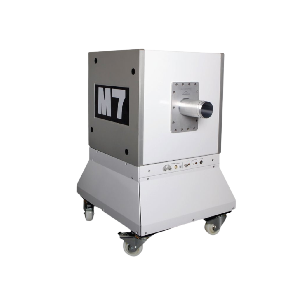
The Aspect Imaging M-Series
In this last section I wanted to focus on the compact, cost effective M-Series MRI systems and the variety of applications it can handle to give a better idea of its immense functionality across disciplines.
Morphology
First, The M-Series systems provide 2D and 3D anatomical and morphological images for in vivo assessment of a variety of organs, tissues, and disease states. This includes inflammation, metabolic disorders and developmental biology.


Cancer
In studying cancer, the versatile M-Series systems allows tumors to be easily detected, visualized, and quantified. The unique characteristics of tumors provide a strong contrast from normal healthy tissue on specific MR images. The M-series systems have even been shown by some groups to provide enhanced tumor visualization compared to higher field systems (see image below).
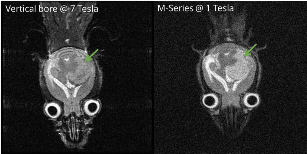
The large gliobastoma tumor (see green arrow) is easier to distinguish with the 1T
M-Series over this 7T system
Neurology
With the M-Series systems anatomical images are easily acquired on the brain and spinal cord. Specific MR images can be acquired to highlight areas of inflammation which are commonly important in neurological disorders and specific anatomical structures. Further, contrast agents may enhance the functional and/or molecular information obtained from these images. Some specific areas of interest include stroke, inflammation and epilepsy.
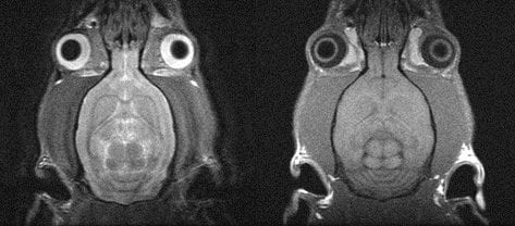
A detailed T1 weighted and T2 weighted image of a rat brain
Contrast Agents
Some contrast agents, specifically Gadolinium based agents, provide a stronger signal in low field systems when compared to higher field systems. The benefit here is increased sensitivity to lower concentrations of contrast agents. This area has been studied greatly, but if you’d like more information about this phenomenon please consider reading the Geninatti-Crich et al. paper: MRI of cells and mice at 1 and 7 Tesla with Gd-Targeting Agents: when low field is better!
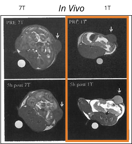
5 hours after injection of a Gadolinium contrast
agent, there is a much greater enhancement in
the lower field MRI (1T) than high (7T)
Other features and add-ons
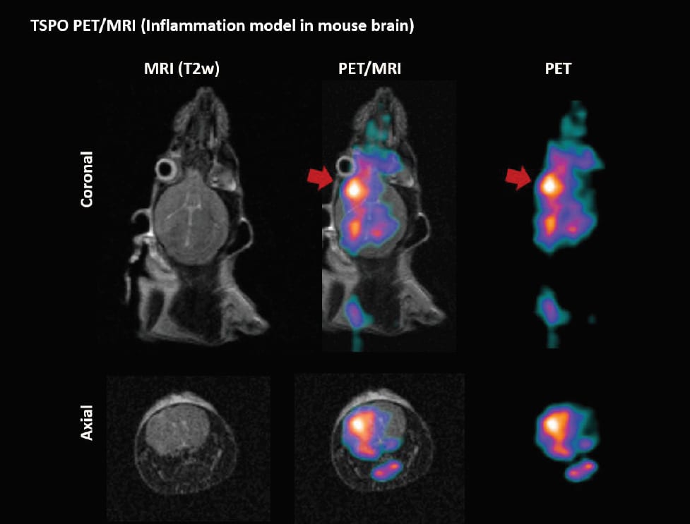
PET/MRI
A PET insert is available for the M-series system for simultaneous PET/MRI images within one system. The 3D anatomical information obtained from the MR image fully complements the highly sensitive PET image in detecting functional information or abnormalities and early disease, helping researchers be able to localize them to specific tissues.
VivoFuse
VivoFuse is both a hardware and software add-on allowing for the co-registration of 3D tomographic optical (BLI/FLI) images with 3D MR images. Combined, the fused data provides the value of both molecular and soft-tissue imaging on the same animal model. The co-registered images allow researchers to confirm that a structure identified on the MR image is a tumor based on the localization of the optical signal to the same region. These combined images provide valuable information to be used when assessing tumor growth, as well as the efficacy of any therapeutic approach.

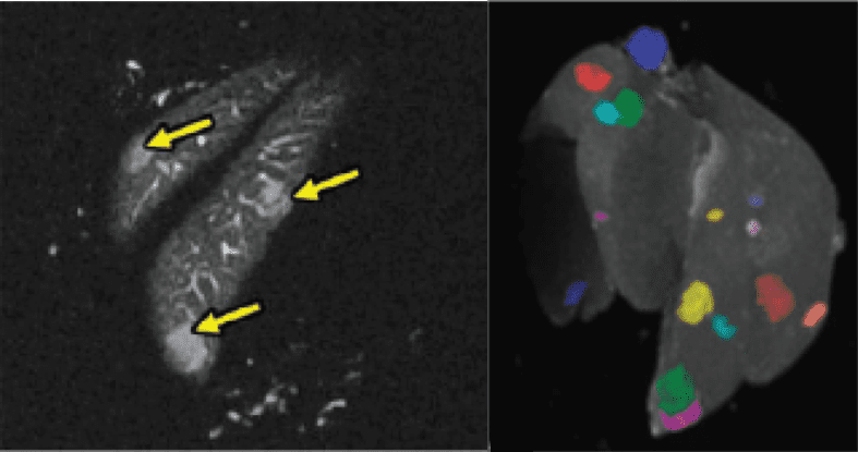
Ex Vivo Imaging Insert
Pre-clinical ex vivo imaging focuses mostly on MR-based histology. In this technique very high-resolution images are acquired on a formalin fixed ex vivo sample. These 3D whole organ images may be used for toxicology studies to help detect, localize, and quantify lesion number and size (see image below). The resulting images are best used to guide the preparation of tissue sections for conventional histological analysis to confirm the pathology associated with the lesions.
Conclusion
I hope after reading this you have come to realize that few applications today need high-field MRI to assist in answering a specific research question. These larger systems, more often than not, are overkill for what a researcher needs to answer their specific question. Smaller 1T systems like the M-series systems offer users a cost-effective option and often open up space for those who absolutely need the high-field systems.
If you have any questions about the M-series systems and their feasibility for your study feel free to reach out to one of our applications specialists at sales@scintica.com or visit the M-series systems web page here.
Keywords: Magnetic resonance imaging, MRI, mouse, rat, rodent, small animal, imaging, contrast agents, low-field, morphology, anatomy, cancer, tumor, PET insert, neuro, neurobiology, contrast agent, in vivo, ex vivo, 1-tesla, 1T, FAQ, frequently asked questions, review.
