Investigating mechanisms driving acute kidney injury arising from heart surgery and sepsis
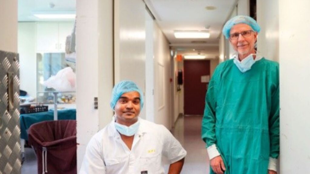
A team of researchers and clinicians at the Florey Institute of Neuroscience and Mental Health are investigating ways to reduce the incidence of kidney injury resulting from heart surgery and sepsis.
Acute kidney injury is when our kidneys suddenly stop working. This happens in up to 30% of patients after heart surgery. Up to 50% of patients who acquire severe infections (‘sepsis’) also develop acute kidney injury. Acute kidney injury leads to a greater chance of dying in hospital, longer stays in hospital, and increased healthcare costs. There are no therapies to prevent kidney injury after heart surgery or sepsis due to our poor understanding of its causes, and the inability to assess the risk of developing kidney injury before it is too late.
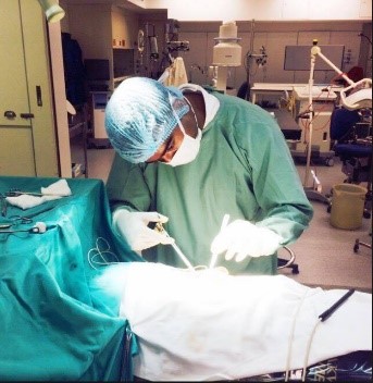
Using clinically relevant sheep models, Dr Yugeesh Lankadeva and colleagues have showed that reduced microvascular blood flow and oxygen levels within the inner part of the kidney (the ‘renal medulla’) is a common pathological feature, which may be driving acute kidney injury after heart surgery and during sepsis. Dr Lankadeva and colleagues have also demonstrated that it is possible to reliably estimate oxygen levels within the renal medulla by measuring oxygen levels within the bladder urine. The placement of a catheter in the bladder is routine clinical practice in patients undergoing heart surgery in operating theatres and patients treated for sepsis in intensive care units. Thus, monitoring bladder urine oxygen levels in such patients provides a relatively non-invasive technique for the early detection of those at risk of developing acute kidney injury. Dr Lankadeva’s recent findings also indicates that it is possible to modify existing therapeutic strategies to improve microvascular blood flow and oxygen levels to the renal medulla, which may reduce the incidence of acute kidney injury arising from heart surgery and sepsis. The development of techniques to simultaneously monitor microvascular perfusion (Oxford Optronix OxyFloTM) and oxygen levels (Oxford Optronix OxyliteTM) in anaesthetized and non-anaesthetized animals provides a very powerful and unique tool to investigate the causes of organ failure arising from heart surgery and sepsis.
References:
- Lankadeva YR, Cochrane AD, Marino B, Iguchi N, Hood SG, Bellomo R, May CN, Evans RG, Strategies that improve renal medullary oxygenation during experimental cardiopulmonary bypass may mitigate postoperative acute kidney injury, Kidney International; 2019, 95:1338-1346
- Iguchi N, Kosaka J, Booth LC, Iguchi Y, Evans RG, Bellomo R, May CN, LankadevaYR. Renal perfusion, oxygenation, and sympathetic nerve activity during volatile or intravenous general anaesthesia in sheep, British Journal of Anaesthesia, 2019 122, 342-349
- LankadevaYR, Kosaka J, Iguchi N, Evans RG, Booth LC, Bellomo R, May CN. Effects of Fluid Bolus Therapy on Renal Perfusion, Oxygenation, and Function in Early Experimental Septic Kidney Injury, Critical Care Medicine, 2019, 47 (1): e36-e43
- Lankadeva YR, Kosaka J, Evans RG, Bellomo R, May CN, Urinary oxygenation as a surrogate measure of medullary oxygenation during angiotensin II therapy in septic acute kidney injury, Critical Care Medicine, 2018, 46 (1): e41-e48
- Lankadeva YR, Kosaka J, Evans RG, Bailey SR, Bellomo R, May CN, Intra-renal and urinary oxygenation during norepinephrine resuscitation in ovine septic acute kidney injury, Kidney International, 2016, 90 (1): 100-108