(March 14, 2024) Webinar: Validation of DEXA for Longitudinal Quantification of Tumor Burden in A Murine Model of Pancreatic Ductal Adenocarcinoma
(March 14, 2024) Webinar: Validation of DEXA for Longitudinal Quantification of Tumor Burden

Selina has been working in intravital imaging research field since 2014. She received Ph.D. in Graduate School of Nanoscience and Technology from KAIST (Korea Advanced Institute of Science and Technology) in 2020. Dr. Ahn has focused her research on understanding in vivo multi-phase spatiotemporal cellular dynamics of transplanted HSPCs in bone marrow during early engraftment using custom-designed video-rate intravital confocal microscopy system. In addition, she has extensive in vivo research experiences on comprehensive 3D spatial/temporal visualization and imaging analysis of various organs such as lymph node, lung, liver, spleen, brain… etc. As an application specialist in IVIM Technology, she provides hands-on practical supports for researchers in various research fields to take full advantage of intravital imaging techniques to maximize their research performance.
(March 14, 2024) Webinar: Validation of DEXA for Longitudinal Quantification of Tumor Burden
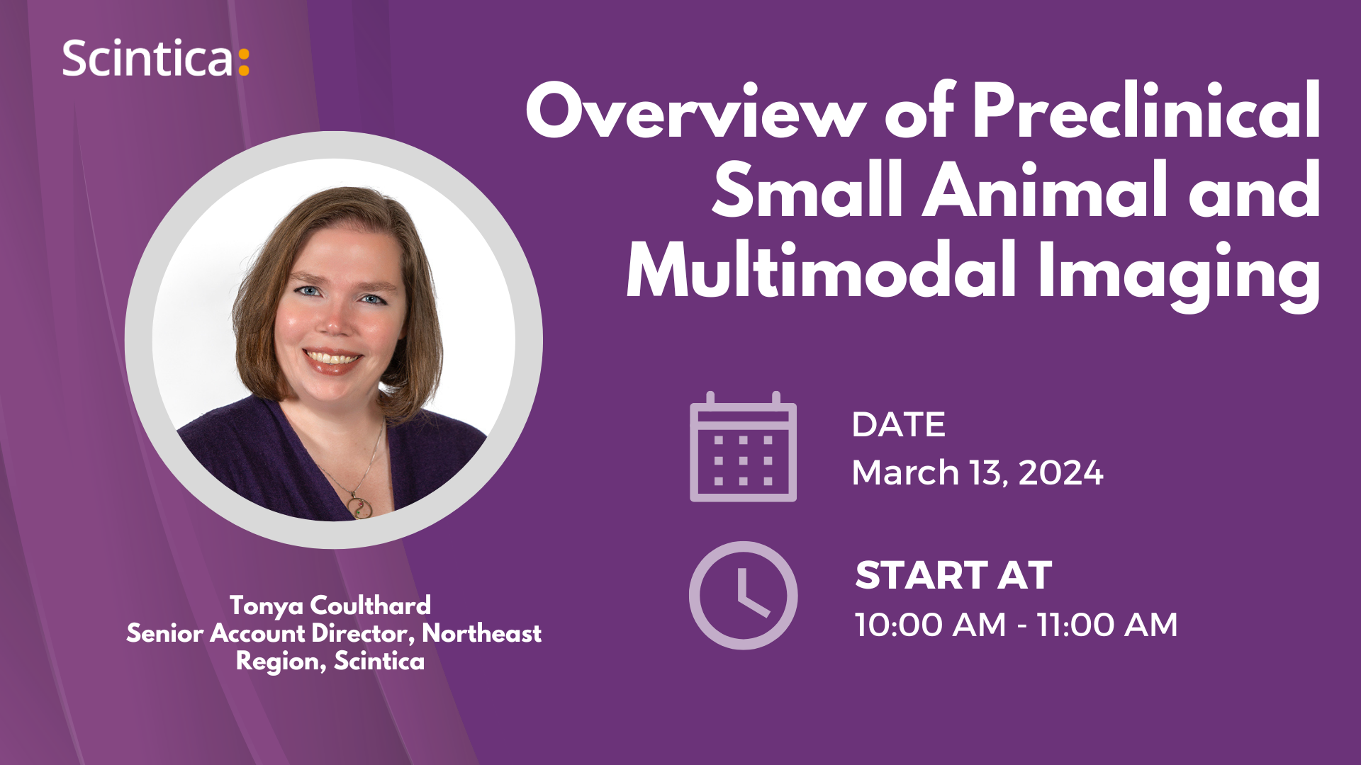
(March 13, 2024) Overview of Preclinical Small Animal and Multimodal Imaging Overview:In this
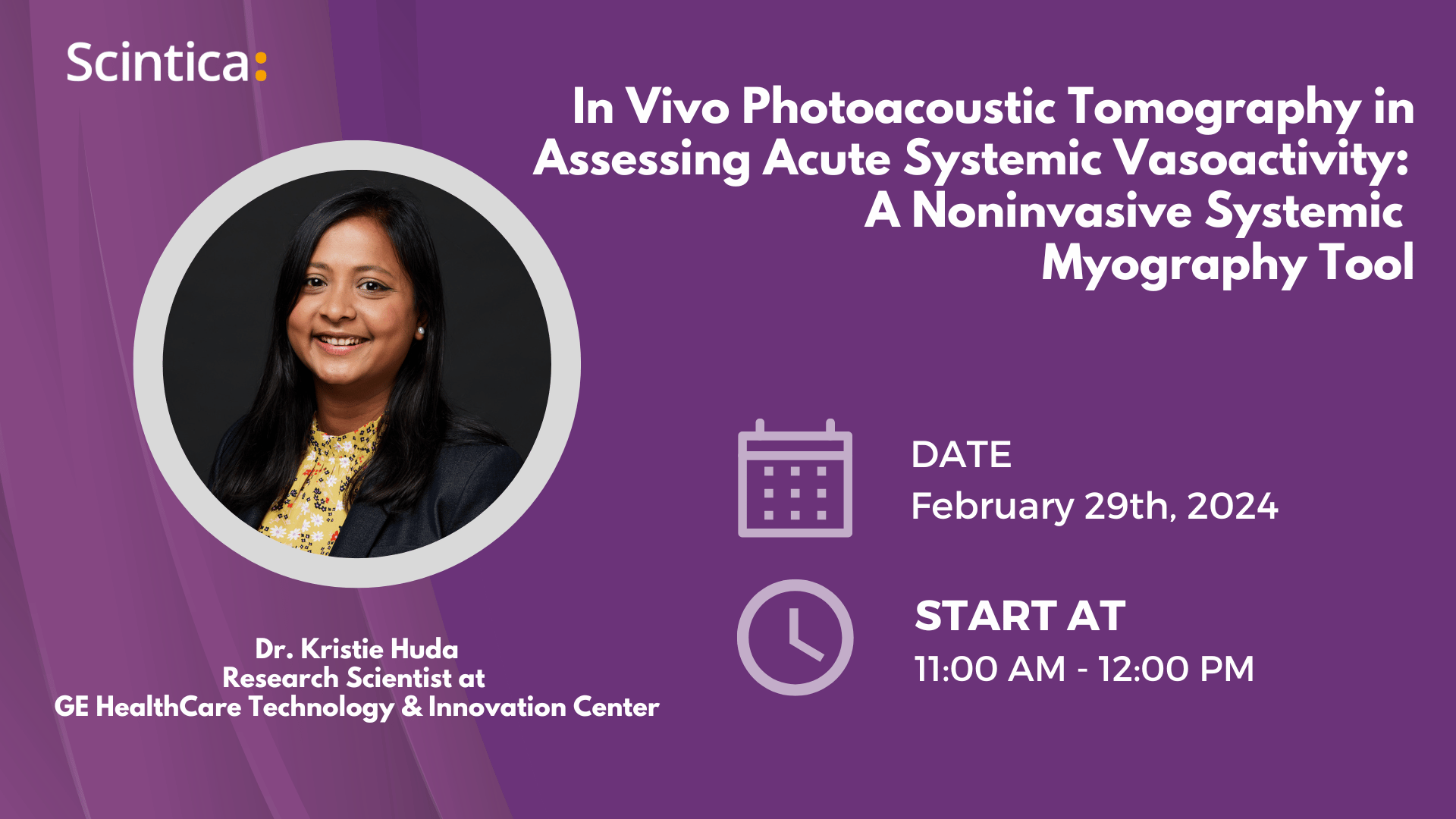
(February 29, 2024) Webinar: In Vivo Photoacoustic Tomography in Assessing Acute Systemic Vasoactivity:
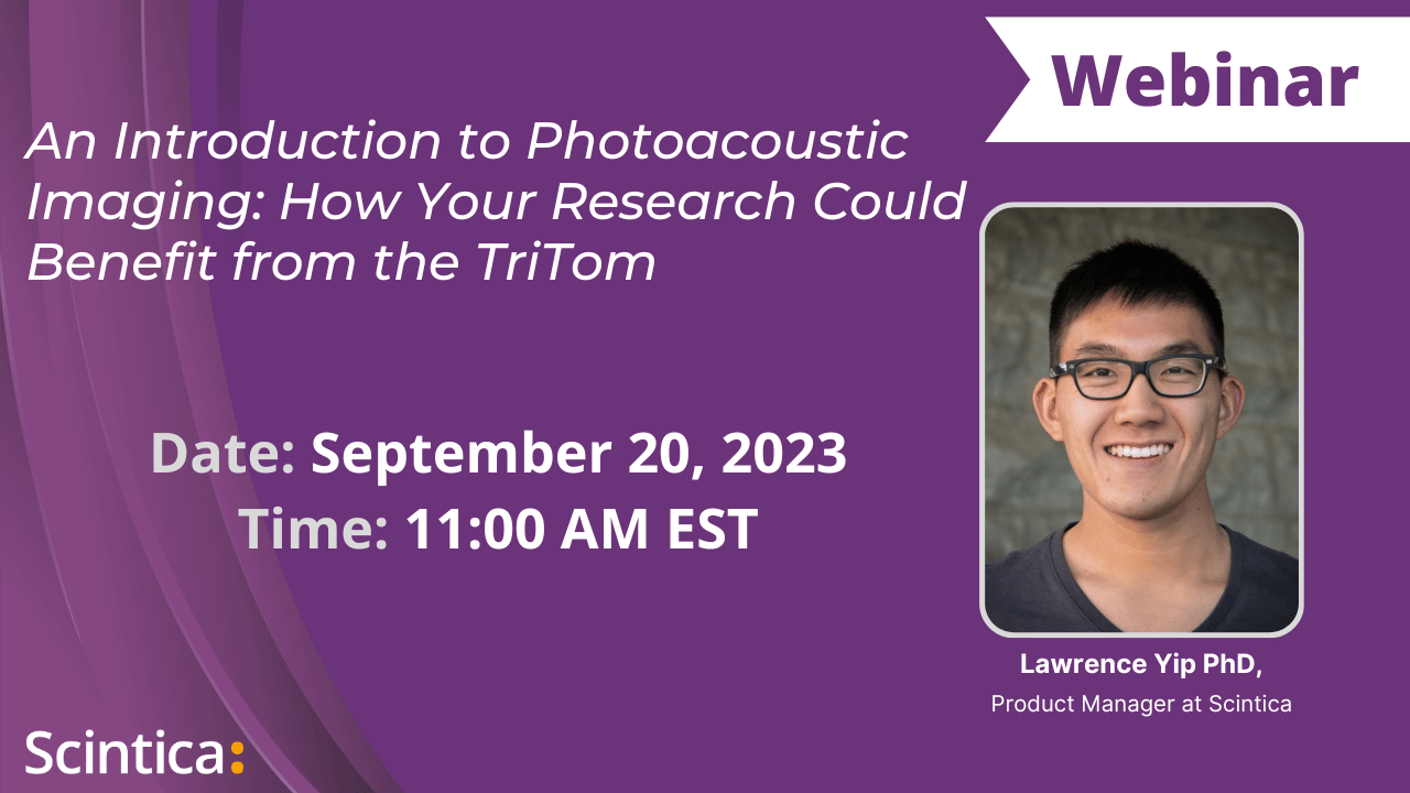
(September 20, 2023) Webinar: An Introduction to Photoacoustic Imaging Overview: Dr. Lawrence Yip
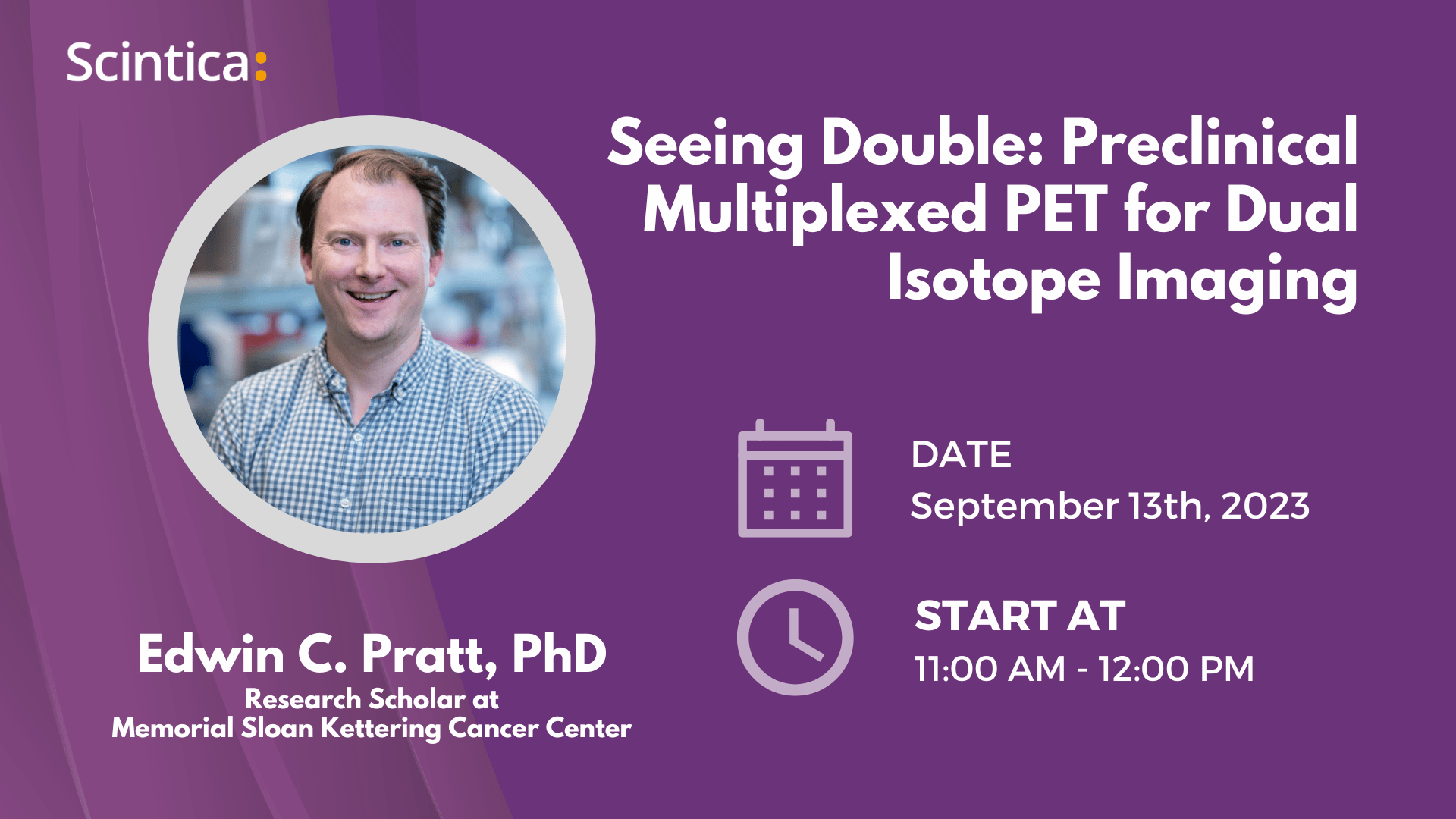
(September 13, 2023) Webinar: Seeing Double: Preclinical Multiplexed PET for Dual Isotope Imaging
(June 29, 2023) Webinar: Designer and Targeted Contrast Agent for Photoacoustic Imaging Overview:The