


Clinical vs. Preclinical Ultrasound: Why Frequency Matters
Clinical vs. Preclinical Ultrasound: Why Frequency Matters View Paper Here Our Ultrasound SystemProspect

Scintica Morning Coffee Chat Episode 1: DXA & Ultrasound
https://youtu.be/KfbEW_b5Gpg?si=2xXwoxw-DuGtjq77 Join us on our first episode of the Scintica Morning Coffee Chat. This
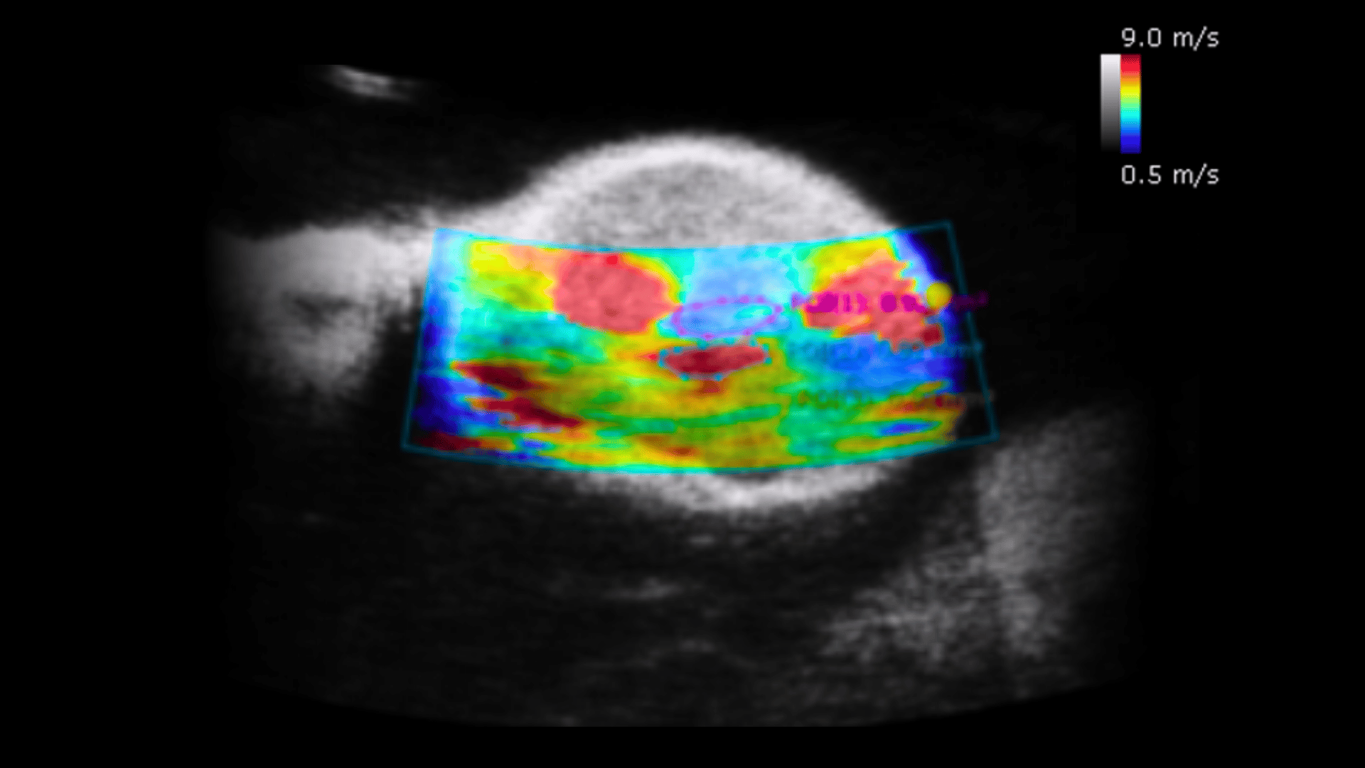
Science Corner: Utilizing Ultrasound Imaging & Shear Wave Elastography to Evaluate Therapy Response of Tumors at an Early Stage
Utilizing Ultrasound Imaging & Shear Wave Elastography to Evaluate Therapy Response of Tumors

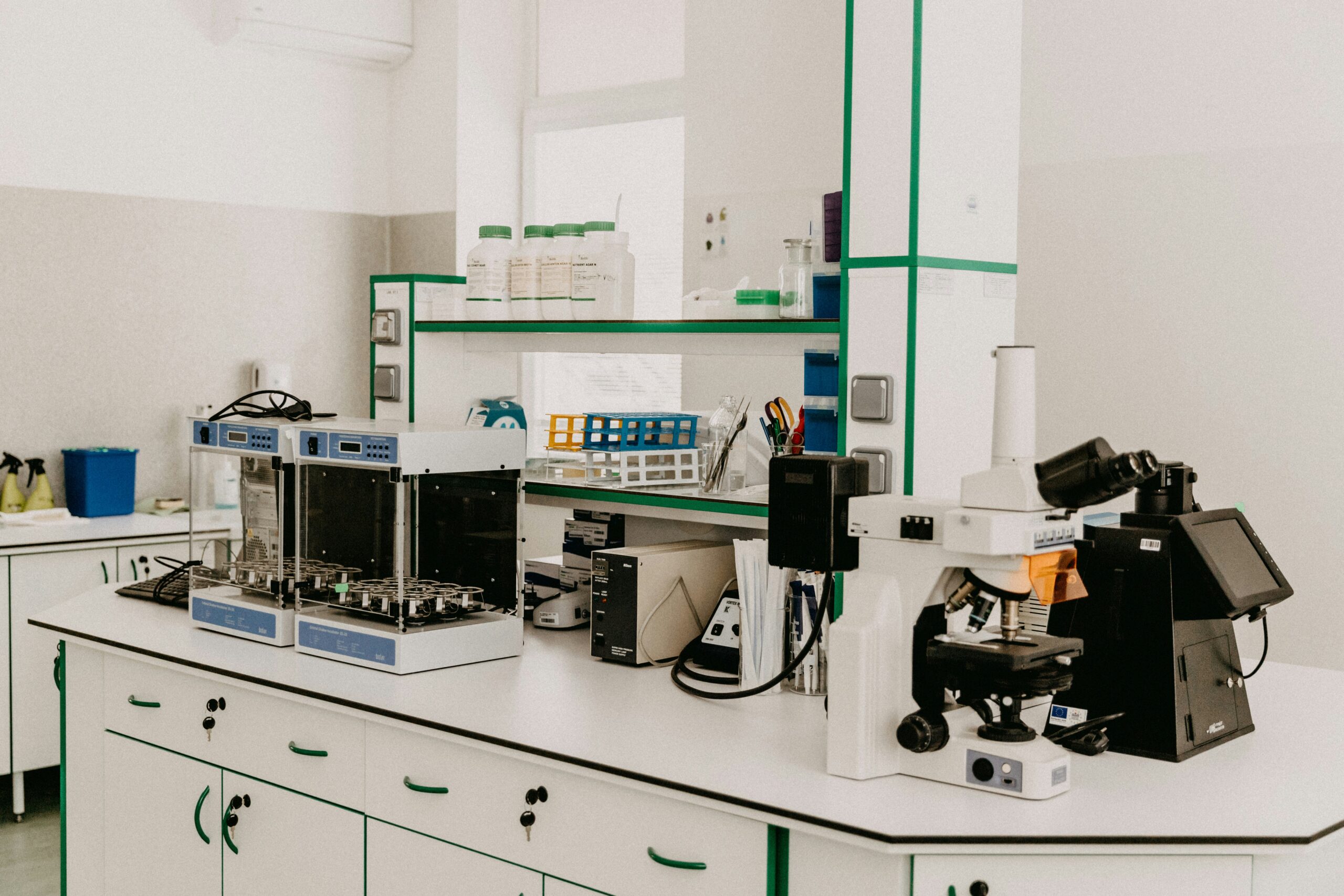
Science Corner: Ultrasound Imaging at Dr. Prasanth Chelikani’s Lab
Innovations in Hypertension and Stroke Research Enhanced by Ultrasound Imaging Dr. Prasanth Chelikani’s
Science Corner: Innovations in Radiolabelling Techniques
Innovations in Radiolabelling Techniques and Radioimmunotherapy Enhanced by Ultrasound Imaging Dr. Jason Holland’s
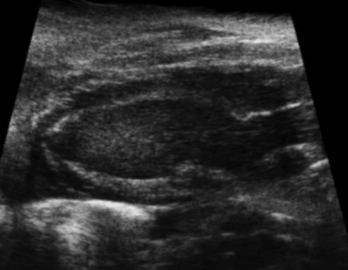
Allogeneic Expanded Human Peripheral NK Cells Control Prostate Cancer Growth in a Preclinical Mouse Model of Castration-Resistant Prostate Cancer
System Used:Prospect T1New Model Available Fangming Wang, Xuejiao Dong, Jing Wang, Feiya Yang,