(August 26, 2021) Webinar #4 Live Virtual Demonstration of the IVM System
Overview:
Welcome to the final 4-part series of the IVIM Intravital Microscopy. Using available models, we walked attendees through the system’s workflow as a user and provide in-depth information on setting up an animal for imaging, acquiring microscopic images using the software, and applying some post-processing techniques to obtain quantifiable results. Participants had the opportunity to ask questions throughout the webinar, which the team answered in real-time. You will learn about: Molecular imaging methods in brief Intravital Microscopy for real-time visualization of live tissues and organs IVM-MS, a new rapid all-in-one technology for Intravital Microscopy Animal models and applications of the technique After attending this webinar, attendees will understand the workflow of the IVM system, its hardware and software components, and its capabilities to acquire quantifiable microscopic images from dynamic processes occurring in live animals.
Complete the form below to get the password
Access is immediate
About the Speaker (s)
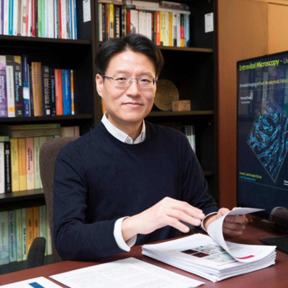
Dr.Pilhan Kim
Associate Professor, Korea Advanced Institute of Science and Technology, Daejeon, Korea
CEO, IVIM Technology, Inc., Daejeon, Korea
Pilhan Kim received his bachelor’s and PhD degree in Electrical Engineering from Seoul National University (Korea) in 2000 and 2005, respectively. From 2005 to 2010, he worked as a postdoctoral research fellow at Harvard Medical School (Boston, USA) with a cross-disciplinary postdoctoral fellowship from Human Frontier Science Program (HFSP). In 2010, he joined the Advanced Institute of Science and Technology (KAIST) where he is currently a tenured Associate Professor at the Graduate School of Nanoscience and Technology. His main research interests focus on systemic cellular-level visualization of various preclinical models to investigate complex pathophysiology of human disease, leading to the development of an advanced in vivo cellular imaging technology based on an ultrafast laser-scanning intravital microscopy system.