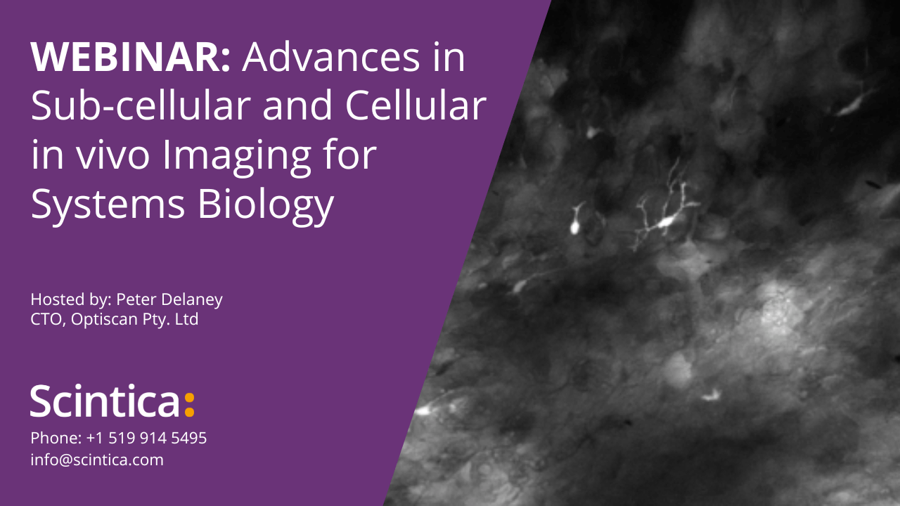
WEBINAR Advances in Sub-cellular and Cellular In Vivo Imaging for Systems Biology
Peter Delaney reviews current trends and technological advancements in the field of in vivo fluorescence imaging, with a focus on new applications and capabilities as realized by confocal fluorescence microscopy and 3D automated optical sectioning.
In vivo fluorescence microscopy is advancing rapidly with the development of new imaging modalities and fluorescence labeling agents. It is now possible to image morphological, molecular, genetic, pathological and physiological events in the living animal. Laser scanning microscopes have advanced quickly, and even miniaturized confocal microscopes of endoscopic proportions can now create 3D observations at the cellular and sub-cellular level in vivo. This has led to a proliferation of techniques facilitating the transition of therapeutic research, bench-to-bedside, through direct in vivo observation of systems biologic processes.
In this exclusive webinar sponsored by Scintica Instrumentation, Peter Delaney discusses unique combinations of advanced imaging instrumentation, fluorescent probes and key applications in terms of their capabilities, limitations and possibilities. Discussion centers on the importance of sub-cellular and cellular observations in vivo, often producing results counter to in vitro cellular behavior, and therefore streamlining research paths. In addition, he shares a variety of sample images, demonstrating novel capabilities in the world of high-resolution in vivo confocal endomicroscopy.
Key topics covered during this webinar include…
- fundamentals of confocal fluorescence microscopy
- generating 3D images using automated Z-stacking
- choosing the right fluorophore/dye for your research application
- planning your imaging experiments with the end (result) in mind
Timestamps:
2:55 – Why is In Vivo microscopy important
8:18 – Overview of current In Vivo microscope technologies
17:24 – 3D image of visceral adipose tissue
25:26 – Explanation of miniaturized confocal microscopy
27:21 – The ViewnVivo system
28:23 – Illustrative examples of the ViewnVivo’s capabilities
44:08 – Bundled fibre vs point scanning technology
47:26 – Where does whole body fluorescence imaging fit into the mix
48:50 – Comments on 3D image generation
51:07 – Comments on the laser technology of the ViewnVivo
55:04 – Comments on depth and dyes for ViewnVivo
About the Speaker (s)
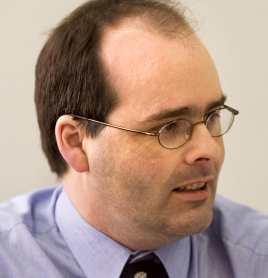
Peter Delaney, CTO
Optiscan Pty. Ltd
Mr. Peter Delaney completed a science degree with honors in Pharmacology at Monash University in 1989. He has played a major role in the refinement of the fibre optic approach to produce a commercial instrument which received an R&D 100 Award in 1991. In 1993, Mr Delaney received the Victorian Young Achiever Award (Science and Technology) for his development of the company strategy and infrastructure, and in 2007 was awarded a prestigious ATSE Clunes Ross award for excellence in the innovation and commercialization of scientific endeavors.